
What Is a Venous Malformation of the Sciatic Nerve?
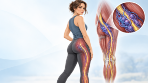
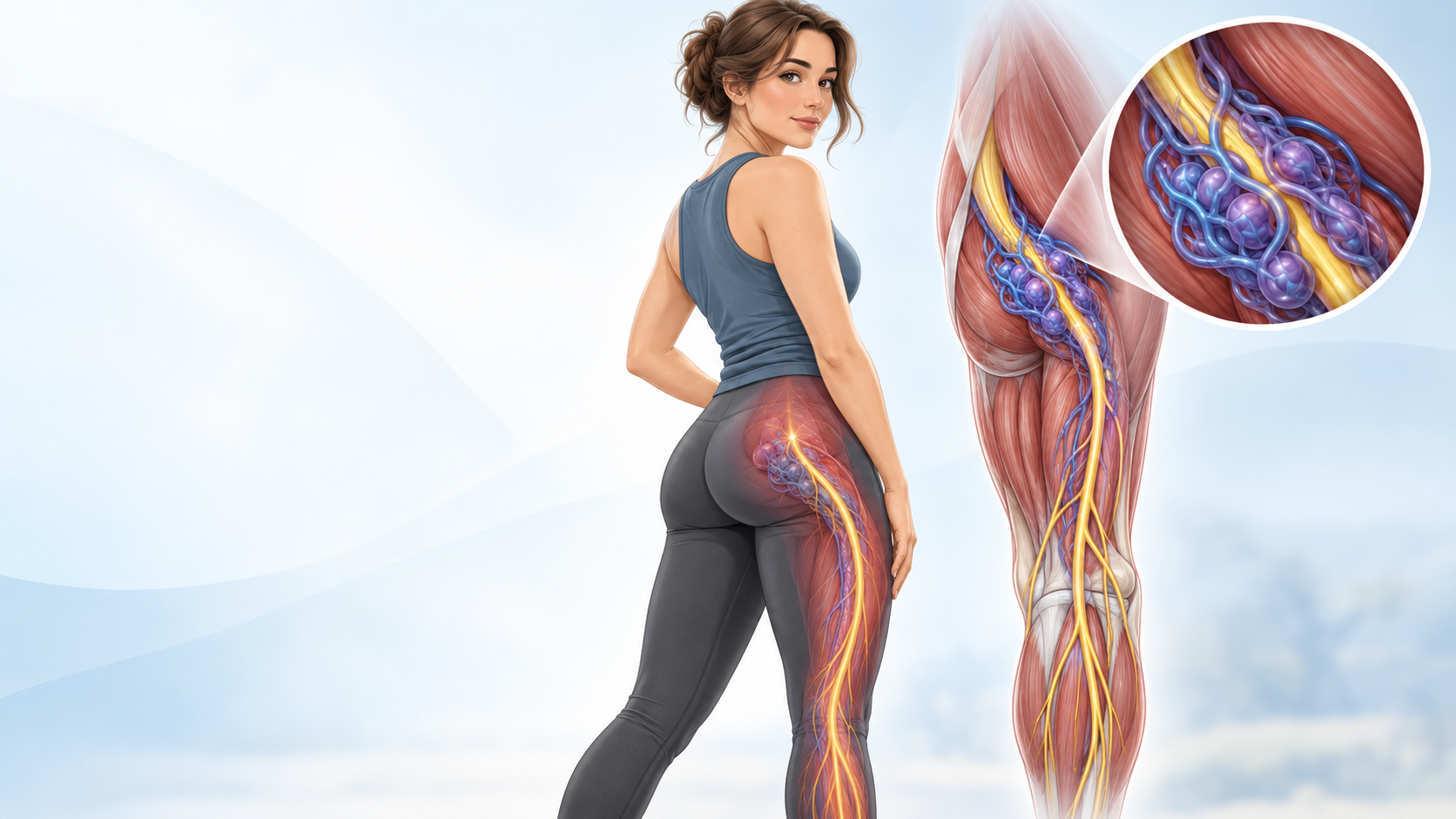
A venous malformation of the sciatic nerve is a rare condition in which abnormal veins develop inside, around, or very close to the sciatic nerve.
A venous malformation, often shortened to VM, is a group of veins that did not develop normally. Venous malformations are considered slow-flow vascular malformations. They are not cancer, and they are different from vascular tumors such as hemangiomas. Venous malformations usually develop before birth, even though symptoms may not appear until childhood, adolescence, or adulthood.
Most venous malformations occur in soft tissue. Involvement of a major peripheral nerve is much less common.
When a VM develops along the sciatic nerve, it can irritate, compress, surround, or sometimes grow within the tissues of the nerve itself. This may cause symptoms that look very similar to ordinary sciatica.
However, the cause is different.
Instead of a spinal disc pressing on a nerve root, the problem may be abnormal veins affecting the sciatic nerve farther down the leg.
Published medical reports have documented venous and other vascular malformations involving the sciatic nerve and causing sciatic neuropathy.
Can a Venous Malformation Follow the Sciatic Nerve Into the Peroneal Nerve?
Yes. A venous malformation involving the sciatic nerve may sometimes extend along the nerve toward its common peroneal branch, also called the common fibular nerve.
It is more accurate to say that the malformation extends along or involves these connected nerve structures rather than saying that it “travels” down the nerve.
Understanding the anatomy explains why this can happen.
The sciatic nerve is the largest nerve in the body. It travels from the lower spine and pelvis, through the buttock, and down the back of the thigh.
Near the knee, the sciatic nerve divides into two major branches:
Sciatic nerve → Tibial nerve + Common peroneal (common fibular) nerve
The common peroneal nerve then passes toward the outside of the knee, near the head of the fibula, before dividing again into the superficial and deep peroneal nerves.
Because the common peroneal nerve is directly connected to the sciatic nerve, a vascular abnormality affecting the sciatic nerve can potentially continue along tissues associated with the peroneal division.
MRI research has also shown that venous malformations in the thigh can infiltrate surrounding muscles as well as the sciatic nerve.
This type of involvement is uncommon, so it should usually be evaluated by doctors experienced with both vascular anomalies and peripheral nerves.
What Does “Intraneural Venous Malformation” Mean?
Doctors may use several terms when describing the relationship between a venous malformation and a nerve.
Extraneural
Extra means outside.
An extraneural venous malformation sits outside the nerve but may push against it.
Imagine a water-filled balloon pressing against an electrical cable. The cable itself may be normal, but pressure from the balloon can interfere with how it works.
Perineural
A perineural malformation grows around or alongside the nerve.
It may surround part of the nerve without growing deeply between its individual nerve fibers.
Intraneural
Intra means inside.
An intraneural venous malformation involves structures inside the nerve covering itself.
This is especially important because a nerve is not one solid wire. It contains many small bundles of nerve fibers called fascicles.
If abnormal veins grow between or around these fascicles, treatment becomes much more difficult because damaging the abnormal veins could also damage healthy nerve fibers.
Intraneural vascular abnormalities are rare. A medical review identified only a small number of reported peripheral nerve cases, illustrating how unusual these lesions are.
Why Can a Sciatic Nerve Venous Malformation Cause Pain?
A venous malformation can affect a nerve in several ways.
1. Pressure on the nerve
Abnormal veins can become enlarged as they fill with blood.
If the veins surround the sciatic nerve, increased pressure can irritate the nerve.
This can cause:
- aching pain
- shooting pain
- burning
- tingling
- numbness
- electric-shock sensations
Johns Hopkins notes that a venous malformation near a nerve can damage the nerve and cause pain or reduced function.
2. Swelling inside or around the nerve
Blood moves slowly through a venous malformation.
The malformation can therefore change in size or pressure at different times. Some patients notice more pain or swelling after activity or after being in certain positions.
Depending on its location, sitting may place extra pressure on a VM near the sciatic nerve.
3. Small clots inside the malformation
Slow-moving blood can sometimes form small clots inside a venous malformation.
These clots can cause a sudden increase in tenderness, swelling, or pain.
Over time, some clots can calcify and form small hard structures called phleboliths.
4. Direct involvement of nerve fibers
If the VM is truly intraneural, the abnormal vessels may affect the internal structure of the nerve.
This can interfere with the electrical signals traveling between the spinal cord, leg, ankle, and foot.
Symptoms of a Venous Malformation Affecting the Sciatic Nerve
Symptoms depend on exactly where the malformation is located and how much of the nerve is involved.
A person may experience:
- deep buttock pain
- pain in the back of the thigh
- aching or pressure in the leg
- burning pain
- shooting or electric pain
- numbness
- pins and needles
- abnormal skin sensation
- leg weakness
- muscle fatigue
- pain while sitting
- pain during or after exercise
- swelling of the thigh or leg
- tenderness over the malformation
- difficulty walking
Some people may have only pain at first.
Others develop neurological symptoms as nerve involvement becomes more significant.
Published cases of vascular malformations affecting the sciatic nerve have included radiating leg pain and other signs of sciatic neuropathy.
What Happens When the Common Peroneal Nerve Is Involved?
Peroneal nerve involvement can produce a more specific group of symptoms.
The common peroneal nerve, also known as the common fibular nerve, controls important muscles in the lower leg and carries sensation from parts of the lower leg and foot.
When it is irritated or damaged, symptoms can include:
Numbness or tingling
A person may notice altered sensation along:
- the outside of the lower leg
- the top of the foot
- parts of the toes
Exactly where the numbness occurs depends on which branch of the peroneal nerve is affected.
Weakness lifting the foot
The deep peroneal nerve helps control muscles that lift the foot upward.
Weakness may make it difficult to:
- lift the front of the foot
- walk on the heels
- clear the toes from the ground while walking
Foot drop
More severe peroneal nerve dysfunction can lead to foot drop.
Foot drop means the front of the foot cannot be lifted normally.
A person may compensate by lifting the knee higher while walking to prevent the toes from dragging.
New or worsening foot drop is an important neurological symptom and should receive prompt medical evaluation.
Ankle weakness
A person may also have difficulty moving the foot outward because other branches of the peroneal nerve control the muscles responsible for eversion.
Why Is This Condition Sometimes Mistaken for Ordinary Sciatica?
Sciatica is extremely common.
A venous malformation of the sciatic nerve is extremely uncommon.
Because of this difference, doctors usually look for common causes first.
Most typical sciatica originates near the spine, where a lumbar disc, arthritis, or narrowing around a nerve root causes irritation.
A venous malformation can produce a similar pattern of pain farther along the sciatic nerve.
This is called an extraspinal cause of sciatic neuropathy.
In a published Mayo Clinic case series, four people had radiating leg pain caused by vascular abnormalities affecting the sciatic nerve. The authors recommended high-resolution imaging of the sciatic nerve when symptoms suggest sciatic neuropathy but standard spinal evaluation does not explain the problem.
This does not mean everyone with unexplained sciatica has a vascular malformation. These lesions remain very rare.
It means that persistent or unusual symptoms sometimes require doctors to look beyond the spine.
How Is a Sciatic Nerve Venous Malformation Diagnosed?
Diagnosis usually requires a combination of:
- medical history
- neurological examination
- vascular examination
- MRI
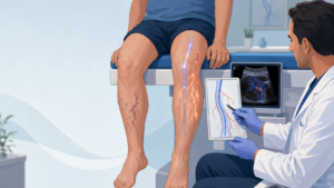
- sometimes ultrasound
- sometimes nerve testing
- sometimes blood tests
Because this is an uncommon condition, diagnosis can require input from more than one specialist.
MRI Is Usually the Most Important Imaging Test
Magnetic resonance imaging, or MRI, is generally the most useful test for defining a venous malformation.
MRI can show:
- the size of the malformation
- its exact location
- whether it involves muscle
- whether it surrounds a nerve
- whether it enters the nerve
- its relationship to the sciatic nerve
- whether it extends toward the tibial or common peroneal nerve
- nearby arteries and veins
Johns Hopkins describes MRI as the best imaging study for defining the size and location of a VM and its relationship to nerves, muscles, arteries, and veins.
When nerve involvement is suspected, doctors may request high-resolution MRI or MR neurography to examine the nerve itself more carefully.
High-resolution MRI is especially helpful before considering surgery because the exact relationship between the abnormal vessels and nerve fascicles matters greatly.
What Can a Venous Malformation Look Like on MRI?
The appearance varies from patient to patient.
Venous malformations often appear very bright on fluid-sensitive MRI sequences such as T2-weighted or STIR images.
MRI may reveal:
- clusters of abnormal venous channels
- enlargement of affected tissues
- extension through several muscles
- involvement around a peripheral nerve
- intraneural involvement
- small clots
- phleboliths
- patterns of contrast enhancement
MRI is also important because the doctor must distinguish a slow-flow venous malformation from a high-flow arteriovenous malformation, or AVM.
These are different conditions and may require different treatment strategies.
Can Ultrasound Detect a Venous Malformation Around a Nerve?
Yes.
Ultrasound, especially high-frequency ultrasound with Doppler, can be useful for examining peripheral nerve vascular abnormalities.
It can help doctors see:
- the nerve
- abnormal veins
- blood flow
- whether a lesion is compressible
- its relationship with surrounding tissue
In a 2024 study of patients evaluated for intraneural vascular abnormalities, ultrasound showed strong agreement with MRI and was considered a useful diagnostic tool.
However, MRI usually provides a better view of the full extent of a deep lesion such as one involving the sciatic nerve.
What Are EMG and Nerve Conduction Studies?
Doctors may also perform:
EMG — electromyography
and
NCS — nerve conduction studies.
These tests measure how well nerves send electrical signals and how muscles respond.
They can help determine whether there is:
- sciatic neuropathy
- peroneal neuropathy
- tibial nerve involvement
- muscle denervation
- nerve damage coming from the spine
However, a normal nerve test does not always completely rule out a problem.
In reported vascular causes of sciatic neuropathy, neurological testing has sometimes been less clear than the MRI findings.
Blood Tests May Be Important in Larger Venous Malformations
Large or deep venous malformations can sometimes develop a condition called localized intravascular coagulopathy, or LIC.
The name sounds complicated, but the basic idea is simple.
Blood moves slowly inside the abnormal veins. This can cause repeated small amounts of clotting inside the malformation.
Doctors may check:
- D-dimer
- fibrinogen
- platelet count
- other clotting tests
Research has found that larger and deeper venous malformations are more likely to show this type of clotting activity.
This is one reason patients with extensive venous malformations may benefit from assessment by a hematologist.
Blood-thinning medicines should not be started without medical advice because treatment depends on the individual patient’s clotting and bleeding risks.
How Is a Venous Malformation of the Sciatic or Peroneal Nerve Treated?
There is no single treatment that is right for every patient.
Treatment depends on:
- where the VM is located
- whether it is inside or outside the nerve
- how much of the nerve is involved
- symptoms
- muscle weakness
- foot drop
- pain severity
- clotting problems
- size of the malformation
- nearby muscles and blood vessels
- previous treatment
The main goals are usually to:
reduce pain, protect nerve function, preserve movement, control the malformation, and avoid causing additional nerve damage.
Observation
Not every venous malformation requires immediate treatment.
If a lesion is stable and causes little or no pain, weakness, or functional difficulty, doctors may recommend monitoring it.
Venous malformations are benign, meaning they are not cancer. Treatment is usually based on symptoms, function, location, and risk rather than simply the presence of the abnormal veins.
Monitoring may include repeat examinations and MRI scans.
Sclerotherapy
Sclerotherapy is one of the most widely used treatments for venous malformations.
An interventional radiologist inserts a needle directly into the abnormal veins using imaging guidance.
A medication called a sclerosant is injected into the malformed veins. It damages the lining of the abnormal vessels, causing them to close and scar down.
Sclerotherapy can reduce:
- pain
- swelling
- pressure
- size of the malformation
Multiple procedures may be needed.
Why nerve involvement makes sclerotherapy more complicated
When a VM is immediately beside or inside the sciatic or peroneal nerve, treatment requires additional caution.
Sclerotherapy causes inflammation and swelling as the treated veins close.
Near a major nerve, doctors must consider whether this temporary swelling or the treatment itself could injure nerve tissue.
There are published reports of sciatic nerve injury after vascular interventions, reinforcing why lesions near major nerves should be treated by teams experienced in complex vascular anomalies.
The question is therefore not simply:
“Can this malformation be sclerosed?”
The more important question is:
“Can the target portion of the malformation be treated without creating an unacceptable risk to the sciatic or peroneal nerve?”
Surgery
Surgery may be considered in selected cases.
Possible surgical goals include:
- removing a well-defined portion of the VM
- relieving pressure on the nerve
- performing neurolysis
- protecting or separating nerve fascicles
- improving pain or function
Neurolysis means carefully freeing a nerve from tissue that is compressing or surrounding it.
A Mayo Clinic series involving rare vascular abnormalities of the sciatic nerve reported improvement in pain following external and limited internal neurolysis in selected patients.
However, surgery becomes particularly difficult when a venous malformation grows between nerve fascicles.
Research on intraneural vascular abnormalities emphasizes that surgical removal can cause nerve injury and that recurrence is possible. The exact compartment of the nerve involved can influence whether safe resection is realistic.
For this reason, complete removal is not always the best goal.
Sometimes preserving nerve function is more important than removing every abnormal vein.
Medication
Medication may be used for several different reasons.
Pain treatment can include medications selected according to whether the pain is inflammatory, muscular, or neuropathic.
Some patients with extensive venous malformations may also receive specialist medications aimed at biological pathways that cause abnormal vascular growth.
For example, sirolimus is sometimes used for complex or extensive vascular malformations. It requires specialist supervision and regular blood monitoring.
Patients with significant localized clotting problems may sometimes require anticoagulant treatment under the guidance of a hematologist.
These medications are not appropriate for every VM.
Why a Multidisciplinary Vascular Anomalies Team Matters
A sciatic nerve venous malformation sits at the intersection of two complicated systems:
the vascular system and the nervous system.
That is why treatment may involve several specialists rather than a single doctor.
Depending on the case, the team may include:
- interventional radiologist
- vascular anomalies specialist
- peripheral nerve surgeon
- neurosurgeon
- orthopedic surgeon
- vascular surgeon
- diagnostic radiologist
- neurologist
- hematologist
- physiotherapist
- pain specialist
- genetic specialist
Major vascular anomaly centers commonly use multidisciplinary teams because treatment often requires several different approaches.
For a lesion directly involving the sciatic or common peroneal nerve, experience with peripheral nerve anatomy is especially important.
What Questions Should MRI Try to Answer?
For a suspected sciatic nerve VM, simply knowing that “a venous malformation is present” is not enough.
Treatment planning benefits from answering more detailed questions:
- Where does the malformation begin?
- How far down the thigh does it extend?
- Does it touch the sciatic nerve or actually enter it?
- Is it inside the epineurium of the nerve?
- Does it involve individual nerve fascicles?
- Does it continue into the tibial nerve?
- Does it continue into the common peroneal nerve?
- Is muscle involved?
- Are there large draining veins?
- Are there phleboliths or areas of thrombosis?
- Is the lesion definitely slow-flow?
- Is there any arterial component?
- Which part of the malformation is responsible for the patient’s symptoms?
- Which portions could potentially be treated safely?
These questions can be especially important before sclerotherapy or surgery.
Sciatic Nerve VM vs. Common Peroneal Nerve Entrapment
Not every patient with peroneal symptoms has a venous malformation inside the peroneal nerve.
The common peroneal nerve is vulnerable to ordinary compression near the outside of the knee, particularly where it curves around the fibular head.
Typical common peroneal nerve compression can also cause:
- numbness
- tingling
- weakness
- foot drop
Therefore, doctors need to determine whether symptoms come from:
- the sciatic nerve
- the common peroneal nerve
- the lumbar spine
- a vascular malformation
- ordinary nerve compression
- or more than one problem at the same time
This is where MRI, neurological examination, and sometimes EMG or nerve conduction testing become valuable.
Other Conditions That Can Look Similar
Doctors may need to distinguish a venous malformation affecting the sciatic nerve from other conditions, including:
- lumbar disc herniation
- lumbar spinal stenosis
- piriformis or deep gluteal syndrome
- schwannoma
- neurofibroma
- intraneural ganglion cyst
- arteriovenous malformation
- vascular tumor
- fibroadipose vascular anomaly (FAVA)
- muscle or tendon injury
- ordinary common peroneal nerve compression
This is one reason a detailed MRI of the actual painful area can matter when symptoms are unusual or standard spinal imaging does not provide an explanation.
Can a Sciatic Nerve Venous Malformation Get Worse Over Time?
It can.
Venous malformations generally grow along with the body. Symptoms may also become more noticeable at different stages of life.
Pain and swelling can change over time, and some VMs enlarge after trauma or during hormonal changes such as puberty or pregnancy.
A lesion affecting a nerve deserves additional attention because prolonged compression or direct nerve involvement can potentially affect nerve function.
Increasing numbness, weakness, or foot drop may therefore be more important than a simple change in the visible size of the malformation.
Does a Venous Malformation Turn Into Cancer?
No. A typical venous malformation is a benign vascular malformation, not a cancer.
It is also not the same thing as a hemangioma.
The two terms were commonly mixed together in the past, which is why older medical records may use words such as:
- venous angioma
- cavernous hemangioma
- intramuscular hemangioma
Modern vascular anomaly classification tries to distinguish vascular tumors from vascular malformations more clearly.
For patients reviewing older medical reports, it may therefore be useful to have the original images reviewed by a vascular anomalies radiologist rather than relying only on the old terminology.
When Should Someone Seek Prompt Medical Attention?
Anyone with a known or suspected sciatic or peroneal nerve venous malformation should contact their medical team promptly if they develop:
- new weakness
- worsening leg weakness
- new foot drop
- rapidly increasing numbness
- difficulty walking that is getting worse
- sudden major increase in pain
- rapidly increasing swelling
- significant redness or warmth
- major change in leg or foot color
Emergency assessment is appropriate for symptoms such as sudden shortness of breath or chest pain, which can indicate a serious blood clot complication from many possible causes.
Loss of bladder or bowel control or numbness around the groin also requires emergency assessment because these symptoms can indicate serious spinal nerve compression rather than a peripheral sciatic nerve problem.
Can Someone Live Normally With a Sciatic Nerve Venous Malformation?
Many people with venous malformations can maintain good function, but the outlook depends heavily on the exact anatomy.
A small VM sitting beside the nerve is very different from a large malformation that grows through muscles and between nerve fascicles.
The most important factors include:
- how much of the nerve is involved
- whether weakness is present
- whether the malformation is stable
- whether recurrent clotting causes pain
- whether treatment can target the VM safely
- whether muscles and joints are also involved
Because intraneural vascular malformations are rare, there is no single outcome that applies to everyone.
Long-term management may sometimes be more realistic than expecting a one-time cure.
Frequently Asked Questions
Is a venous malformation of the sciatic nerve rare?
Yes. Venous malformations themselves are not extremely rare, but direct involvement of a major peripheral nerve such as the sciatic nerve is uncommon. Published medical literature largely consists of small series and individual reports.
Can a venous malformation cause sciatica?
Yes. A vascular malformation involving or compressing the sciatic nerve can produce pain similar to sciatica. However, spinal causes of sciatica are much more common.
Can a sciatic nerve venous malformation extend into the peroneal nerve?
It can. The common peroneal, or common fibular, nerve is one of the two major terminal branches of the sciatic nerve. A malformation involving the sciatic nerve may therefore extend along structures associated with the peroneal division.
Can peroneal nerve involvement cause foot drop?
Yes. Damage to the common or deep peroneal nerve can weaken the muscles responsible for lifting the front of the foot, potentially causing foot drop.
What is the best scan for a sciatic nerve venous malformation?
MRI is usually the main imaging test. High-resolution MRI or MR neurography may provide additional detail about the relationship between the malformation and individual nerve structures. Ultrasound with Doppler can also be useful.
Can an EMG diagnose the venous malformation?
No. An EMG does not show the abnormal veins themselves. It can help determine whether the sciatic or peroneal nerve is functioning abnormally. MRI or vascular ultrasound is needed to visualize the malformation.
Is sclerotherapy safe when a VM touches the sciatic nerve?
Sclerotherapy is commonly used for venous malformations, but treatment close to a major nerve requires careful planning. The possible benefits must be weighed against risks such as swelling and nerve injury. An experienced vascular anomalies interventional radiologist should assess the anatomy before treatment.
Can the venous malformation simply be surgically removed?
Sometimes, but not always. A VM surrounding a nerve may be easier to separate than one growing between nerve fascicles. Intraneural surgery can risk permanent nerve damage, so treatment must be individualized.
Why does the pain sometimes change from day to day?
Blood flow, swelling, physical activity, position, and small clots inside the malformation may all affect symptoms. Pain from nerve irritation can also fluctuate.
Does having a venous malformation mean someone will develop blood clots?
Not necessarily. However, blood can clot locally inside a VM because of slow flow. Large or deep lesions may sometimes develop localized intravascular coagulopathy, which doctors can investigate with blood tests.
Is a venous malformation hereditary?
Most are not inherited. Some uncommon types are related to inherited conditions or specific genetic changes.
The Most Important Takeaway
A venous malformation involving the sciatic nerve and extending toward the common peroneal nerve is rare but medically recognized.
The connection makes anatomical sense because the common peroneal nerve is one of the two main branches of the sciatic nerve.
These lesions may cause more than ordinary leg pain. Depending on the location and degree of nerve involvement, symptoms can include:
sciatic-type pain → tingling or numbness → peroneal sensory changes → muscle weakness → possible foot drop.
The most important part of evaluation is determining exactly where the abnormal veins are in relation to the nerve.
A malformation next to a nerve, one surrounding a nerve, and one growing between nerve fascicles may require very different treatment plans.
MRI—sometimes with high-resolution peripheral nerve imaging—is central to understanding this anatomy. Treatment may involve observation, sclerotherapy, medication, surgery, neurolysis, or a combination of approaches.
Because treatment of abnormal veins can itself place an involved nerve at risk, complex cases are best evaluated by a multidisciplinary vascular anomalies team with expertise in peripheral nerve disease.
Medical Disclaimer
This article is for educational purposes only and does not provide individual medical advice. Venous malformations involving major nerves are uncommon and highly variable. Diagnosis and treatment should be based on a patient’s symptoms, examination, MRI findings, and assessment by appropriately qualified specialists.
Key medical sources used
- International Society for the Study of Vascular Anomalies (ISSVA) classification of vascular anomalies.
- Johns Hopkins Medicine: Venous Malformation.
- Van Gompel JJ et al. Vascular malformations, rare causes of sciatic neuropathy: a case series. Neurosurgery.
- Prasad NK et al. A radiological classification system for intraneural vascular anomalies: assessment of potential for resection with high-resolution MRI.
- Rusu GM et al. Review of peripheral intraneural vascular anomalies.
- Chen W et al. Study of ultrasonography for intraneural vascular anomalies of peripheral nerves.