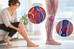

Understanding venous haemodynamics, leg swelling, varicose veins and chronic venous insufficiency.
The problem in your lower leg may actually begin in your pelvis
Many patients experience swollen ankles, heavy legs, painful varicose veins or persistent discomfort in their calves. These symptoms are commonly attributed to poor circulation or faulty valves in the leg veins.
However, sometimes the underlying problem is not in the lower leg at all.
It may be located much higher, deep inside the pelvis.
This is the case with May–Thurner syndrome, a condition in which compression of a major pelvic vein interferes with the normal return of blood from the leg to the heart.
Understanding how this compression changes blood flow—what vascular specialists call venous haemodynamics—helps explain why a problem in the pelvis can produce symptoms all the way down to the ankle.
What is May–Thurner syndrome?
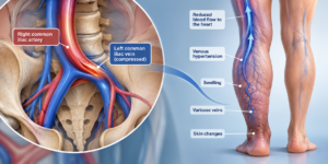
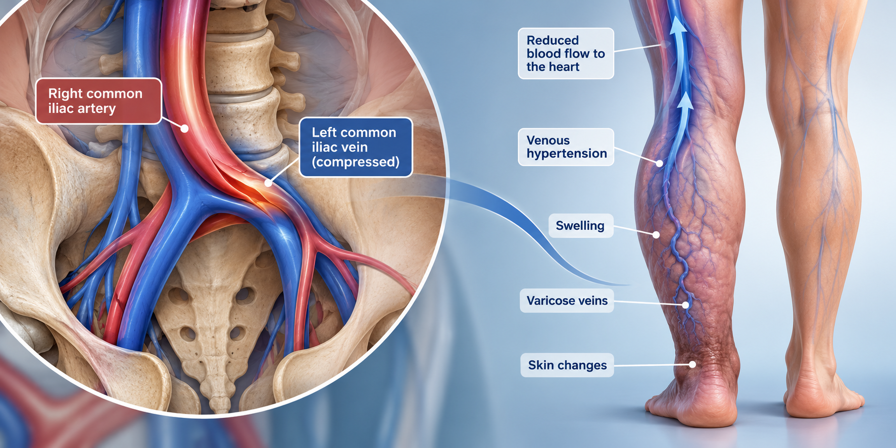
May–Thurner syndrome occurs when the left common iliac vein, one of the main veins draining blood from the left leg, is compressed between the right common iliac artery and the spine.
Unlike arteries, which have relatively thick muscular walls, veins have thinner, more flexible walls. This makes them more susceptible to external compression.
The artery crossing over the vein can narrow it, creating a restriction to venous blood flow.
Over time, repeated arterial pulsations may also cause changes to the inner lining of the vein, including the formation of fibrous tissue that makes the narrowing more persistent.
Importantly, many people have some degree of iliac vein compression without symptoms. May–Thurner becomes clinically important when this narrowing contributes to impaired venous drainage, significant symptoms or blood clots.
Understanding normal blood flow in the legs
To understand the effects of May–Thurner syndrome, we first need to understand how healthy veins work.
Blood travels from the feet and lower legs through deep veins in the calves and thighs. It then passes through the iliac veins in the pelvis before returning to the heart.
Unlike arteries, leg veins do not have a powerful pumping force pushing blood forward.
Instead, venous circulation depends on three essential mechanisms.
1. The calf muscle pump
Each time we walk, the calf muscles contract and squeeze the deep veins, helping propel blood upwards towards the heart.
2. One-way venous valves
Small valves inside many leg veins help prevent blood from falling backwards under the influence of gravity.
3. An unobstructed pathway to the heart
Even when the calf muscles and valves function normally, blood must be able to pass freely through the larger veins in the pelvis.
These three mechanisms work together to maintain efficient venous return.
In a healthy leg, walking normally reduces the pressure within the lower-leg veins. This is one reason movement is beneficial for venous circulation.
But what happens when the main drainage pathway is narrowed?
The haemodynamics of May–Thurner syndrome: why pressure builds up
Imagine a busy motorway carrying traffic towards a city.
If several lanes suddenly merge into one narrow lane, traffic begins to accumulate behind the restriction.
A similar principle applies to venous circulation.
In May–Thurner syndrome, the compressed iliac vein creates increased resistance to blood leaving the affected leg.
The haemodynamic consequences depend on how severe the narrowing is, how much blood needs to pass through it and whether alternative drainage pathways are available.
1. Increased resistance to venous outflow
As the iliac vein becomes narrower, blood has greater difficulty passing through the affected segment.
During exercise, when blood flow from the legs increases, a significant obstruction can become particularly noticeable.
Patients may experience a sensation of tightness, heaviness or bursting discomfort, sometimes called venous claudication.
2. Increased pressure in veins below the obstruction
When venous outflow is restricted, pressure can rise in the venous system below the narrowed segment.
This may involve the veins of the thigh, calf and ankle.
The result is a condition known as venous hypertension, meaning abnormally elevated pressure within the veins.
Unlike high blood pressure measured in an arm artery, venous hypertension refers to increased pressure in the low-pressure venous circulation.
3. Reduced efficiency of the calf muscle pump
Normally, calf muscle contractions empty the deep veins and help reduce venous pressure during walking.
With significant proximal obstruction, blood may not leave the leg efficiently enough to maintain the normal pressure reduction.
This is one mechanism contributing to ambulatory venous hypertension—persistently elevated venous pressure during movement.
The effect can be more pronounced when iliac vein obstruction coexists with damaged or incompetent venous valves.
4. Development of collateral circulation
The body may try to compensate by directing blood through alternative veins, known as collateral veins.
These may develop or enlarge within the pelvis, abdominal wall or other venous pathways.
Collateral circulation can help relieve an obstruction, but it may not fully compensate for the reduced capacity of the main iliac vein.
Some patients remain symptom-free because these alternative pathways provide adequate drainage. Others continue to experience substantial venous congestion.
How does May–Thurner syndrome affect the lower-leg veins?
The consequences of altered venous haemodynamics may eventually become visible in the lower leg.
Swelling of the calf and ankle
One of the common manifestations of impaired venous drainage is swelling.
When venous pressure increases, pressure can also rise within the tiny blood vessels supplying the tissues.
This encourages more fluid to move from the circulation into the surrounding tissues than the lymphatic system can remove.
Patients may notice that the affected ankle or calf becomes progressively swollen throughout the day.
Swelling may worsen after prolonged standing or sitting and improve overnight or when the leg is elevated.
Varicose veins and superficial venous reflux
Varicose veins are enlarged, twisted superficial veins that often result from malfunctioning venous valves.
May–Thurner syndrome does not automatically cause varicose veins. However, significant iliac vein obstruction can contribute to lower-limb venous hypertension and may coexist with, or aggravate, superficial venous reflux.
In some patients, superficial veins may also enlarge as part of the body’s attempt to establish alternative drainage routes.
This distinction is important.
Treating a visible varicose vein does not necessarily correct a significant obstruction higher in the pelvis.
For selected patients with recurrent varicose veins, persistent swelling or symptoms that do not improve after appropriately performed superficial vein treatment, assessment of the pelvic venous outflow may be necessary.
Skin pigmentation and chronic inflammation
When venous hypertension persists, the increased pressure can disturb the microcirculation of the skin.
Small amounts of fluid and blood components may escape from the capillaries into the surrounding tissue.
Over time, this can produce chronic inflammation, brown pigmentation from iron-containing blood pigments, eczema and thickening of the skin.
These changes are often particularly noticeable around the ankle.
Venous leg ulcers
In advanced chronic venous disease, prolonged venous hypertension may contribute to skin breakdown and the formation of venous ulcers.
These wounds commonly occur around the lower leg, particularly near the inner ankle.
Most venous ulcers are associated with venous reflux, previous deep vein thrombosis or a combination of venous abnormalities.
However, iliac vein outflow obstruction can be an additional contributing factor in selected patients, particularly when ulcers are difficult to heal.
May–Thurner syndrome and deep vein thrombosis
Another important consequence of iliac vein compression is an increased risk of deep vein thrombosis (DVT).
Blood flowing slowly through a significantly narrowed vein is more susceptible to clot formation. Chronic compression may also damage the vein lining, further contributing to thrombosis.
A DVT may cause sudden swelling, pain, tenderness and discolouration of the affected leg.
If a clot damages the venous valves or leaves residual obstruction, patients may develop post-thrombotic syndrome, which can cause long-term swelling, pain and skin changes.
Sudden unexplained swelling or pain in one leg requires urgent medical assessment. New breathlessness, chest pain, fainting or coughing blood may indicate a pulmonary embolism and requires emergency medical care.
Why a standard leg ultrasound may not tell the whole story
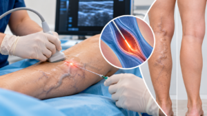
Duplex ultrasound is usually the first investigation for patients with leg swelling, varicose veins or suspected DVT.
It can assess blood flow, detect clots and identify faulty venous valves.
However, the iliac veins are located deep within the pelvis, where routine lower-limb ultrasound may not adequately visualise them.
For patients with symptoms suggesting an outflow obstruction, further investigation may involve CT venography or MR venography.
In selected cases where intervention is being considered, catheter venography and intravascular ultrasound (IVUS) provide more detailed information about the anatomy and severity of the narrowing.
The essential question is not simply whether the iliac vein appears compressed.
The important question is whether that compression is clinically significant and contributing to the patient’s symptoms.
Can treating the pelvic obstruction improve lower-leg symptoms?
Treatment must be individualised.
Patients with incidental iliac vein compression and no symptoms generally do not require treatment.
For patients with mild symptoms, management may include appropriate compression stockings, regular walking, calf muscle exercises and leg elevation.
When significant iliac vein obstruction is associated with substantial symptoms, a vascular specialist may recommend venous stenting.
This minimally invasive procedure places a specially designed stent inside the narrowed iliac vein to help maintain an open pathway for blood returning to the heart.
In appropriately selected patients, restoring venous outflow may reduce swelling, heaviness and venous discomfort. It may also assist the management of some difficult cases of chronic venous disease.
However, venous stenting carries risks, including thrombosis, and requires appropriate follow-up. It does not necessarily reverse existing valve damage or eliminate every varicose vein.
Patients with DVT need specific clot treatment, usually including anticoagulation, with additional procedures considered in selected circumstances.
The key message: healthy leg veins depend on healthy venous outflow
May–Thurner syndrome demonstrates an important principle of vascular medicine:
The condition of the veins in your lower legs cannot always be understood by examining the lower legs alone.
The venous circulation is an interconnected system.
A significant obstruction in the pelvis may influence pressure and drainage throughout the affected limb, contributing to swelling, discomfort and chronic venous disease.
For patients with persistent one-sided leg swelling, recurrent varicose veins, unexplained venous discomfort or difficult-to-heal venous ulcers, it may be appropriate to investigate whether an upstream venous obstruction is contributing to the problem.
Understanding the haemodynamics allows clinicians to look beyond visible veins, identify the relevant causes of venous hypertension and select treatment based on the patient’s complete venous circulation.
Because sometimes the problem you see at the ankle begins much higher in the pelvis.
Further reading
Society for Vascular Surgery – May–Thurner Syndrome
Chronic Venous Insufficiency and Varicose Veins of the Lower Extremities – Clinical Review
International Iliac Venous Stenting Guidelines: Applicability to Australia and New Zealand
Medical disclaimer: This article provides general patient education. It does not replace individual assessment, diagnosis or treatment recommendations from a qualified healthcare professional.