
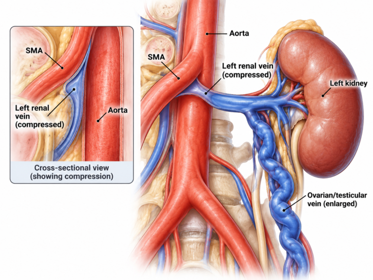
Nutcracker syndrome occurs when the left renal vein becomes compressed between the aorta and the superior mesenteric artery, which can lead to symptoms such as blood in the urine flank pain and pelvic congestion.
Detailed history, examination and vascular ultrasound
tailored to your specific findingsand goals.
experienced team in complex vacular management
conservative and interventional options when appropriate
Nutcracker syndrome occurs when the left renal vein becomes compressed, most commonly between the aorta and the superior mesenteric artery. This compression disrupts venous drainage from the kidney and pelvis, leading to increased venous pressure.
Blood in the urine is one of the most commn symptoms and may be visible or detected on testing. It can be intermittent and may increase with physical activty.
Increased pressure in the left renal vein can cause dull and sharp pain in the left side of the abdomen or flank, which may worsen with prolonged standing or exercise.
In some cases, nutcracker syndrome can contribute to pelvic congestion with symptoms such as pelvic pain, varicose veins in the pelvis or thighs, and a feeling of heaviness.
Symptoms can vary depending on whether the left renal vein is the only one affected or tributaries are also involved. Common symptoms include:

We take a comprehensive approach, combining your history and symptoms with a detailed clinical examination and targeted vascular ultrasound. In selected cases, further imaging such as CT or MR venography may be recommended to confirm the diagnosis and assess the degree of compression.
Nutcracker syndrome can develop gradually as changes in the body’s anatomy affect the way blood drains from the left kidney. In some people, these changes are linked to natural growth during adolescence, significant weight loss or differences in the position of surrounding blood vessels. As the body adapts, smaller veins may widen to provide alternative routes for blood flow. These changes can develop over time, which helps explain why symptoms may appear gradually, fluctuate or remain unnoticed for years. In some younger patients, the condition may improve naturally as the body grows and the anatomy changes.
Treatment depends on the type and severity of your symptoms, We develop a personalised plan based on your diagnosis, lifestyle and goals.
Activity modification
diet modification
Monitoring Urine and Kidney Function
A catheter is inserted into the vein. When it’s in the correct position, a small mesh tube (stent) is put in place in the vein to keep it open
For select cases, we work closely with surgeons to organise surgical procedure reducing compression on the left renal vein.
A member of our team will be in touch soon. Please, allow 24 hours to receive a response to your online form submission.

Something isn’t Clear?
I will be more than happy to answer all of your questions.