

Why Do My Muscles Feel So Tight When My Joints Are So Flexible?
Understanding fascia, myofascial pain and “tissue restriction” in EDS and hypermobility
One of the most confusing symptoms reported by people with hypermobile Ehlers-Danlos syndrome (hEDS) and Hypermobility Spectrum Disorder (HSD) is a feeling of being tight and loose at the same time.
Your joints may move further than they should. Yet the muscles around your neck, shoulders, back, hips or legs may feel incredibly tight.
You might describe your body as:
“My muscles are always tense.”
“It feels like there is a tight band under my skin.”
“I can stretch, but the tight feeling comes straight back.”
“There is a knot that sends pain somewhere else when I press it.”
“It feels as though the tissues do not slide properly.”
These symptoms can be related to muscle guarding, myofascial pain, trigger points and changes in the way layers of connective tissue move over one another.
For some people, these problems are secondary to the joint instability that comes with hypermobility.
Understanding why this happens is important because the answer is not always “stretch more.”
In fact, repeatedly stretching an already hypermobile joint can sometimes make the underlying instability worse.
First, what happens in hypermobility?
In hypermobility, a joint has more movement than usual.
Some people are flexible without having pain or other problems. But in hEDS and HSD, excessive joint movement may be associated with joint instability, recurrent soft-tissue strain and persistent musculoskeletal pain.
In hEDS, joint instability, subluxations, dislocations and soft-tissue injuries can occur with relatively little force. Chronic muscular or myofascial pain is also well recognised.
Think about a tent.
The ropes around the tent help keep it stable. If those ropes become loose, something else has to work harder to stop the tent from wobbling.
Your muscles often do something similar.
When ligaments and other passive supports around a joint do not provide enough stability, the nervous system may ask the surrounding muscles to work harder.
Instead of switching on mainly when you need them, some muscles may remain partly contracted for long periods.
This is called muscle guarding.
Muscle guarding can be useful in the short term because it helps protect an unstable area. But if it continues for hours, days, months or years, the muscles themselves can become painful and tired.
This helps explain the hypermobility paradox:
The joint can be too mobile while the muscles around it feel extremely tight.
Why doesn’t stretching fix the tightness?
Because the problem may not simply be a “short muscle.”
Imagine somebody holding onto a railing because the ground underneath them feels unstable.
Pulling their arm away from the railing does not solve the problem. They may simply grab on again.
A muscle that is constantly working to stabilise a hypermobile joint can behave in a similar way.
You may stretch it and temporarily feel looser. But if the nervous system still believes the joint needs protection, the muscle may tighten again.
This is why treatment for hypermobility-related pain often needs to focus on control, strength and stability, rather than simply increasing flexibility.
Exercise aimed at improving strength, body-position awareness and joint stability is an important part of hEDS management.
Where does fascia fit into this?
Fascia is connective tissue that surrounds and connects structures throughout the body.
It exists around muscles, between muscle groups, around nerves and blood vessels, and in many other areas.
You can think of fascia as thin but strong sheets and layers of tissue that help organise the body’s structures.
Importantly, fascia is not just packaging.
Research shows that deep fascia contains many sensory nerve endings. Some help the brain understand movement and body position. Others are nociceptors—nerve endings that can detect potentially harmful mechanical or chemical changes and contribute to pain.
Fascial layers also normally move or glide over neighbouring structures as we move.
When an area has been injured, overloaded or held under tension for a long time, this movement may become uncomfortable or altered.
That may contribute to the feeling patients describe as a tissue restriction.
Does “tissue restriction” mean your fascia is literally stuck?
Not necessarily.
This is an important distinction.
The term fascial restriction is commonly used to describe an area that feels unusually tight, tender or poorly mobile during examination.
But it does not always mean that two pieces of fascia have physically glued themselves together.
Sometimes there may be true scarring or adhesions. In other situations, the sensation may involve a mixture of increased muscle tension, reduced movement between tissue layers, irritation of sensory nerves, changes in the tissue’s mechanical properties and increased sensitivity of the nervous system.
Researchers are still working out exactly how these factors interact.
So when we talk about a fascial or tissue restriction, we are describing a clinical pattern, rather than claiming that every painful area contains visible scar tissue.
What is myofascial pain?
The word myofascial combines two words:
“Myo” means muscle.
“Fascial” refers to fascia.
Myofascial pain is pain arising from muscles and their related connective tissues.
People may experience aching, pressure, pulling, stiffness, burning or deep tenderness.
One common feature is a myofascial trigger point.
A trigger point is usually described as a very sensitive spot within a tight band of muscle. Pressing it may cause pain at the spot itself or reproduce pain somewhere else.
This second response is called referred pain.
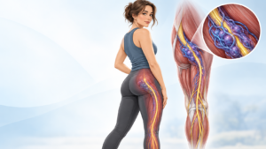
For example, a trigger point around the shoulder blade might produce pain that travels toward the neck or arm.
An international expert consensus identified a taut band, a hypersensitive spot and referred symptoms as important clinical features of trigger points. However, there is still no single blood test, scan or other test that can definitively prove that a trigger point is present.
What is actually happening inside a trigger point?
Scientists are still debating the full answer.
One leading theory starts with overworked muscle fibres.
When a small part of a muscle remains contracted for too long, it continues to use energy.
That sustained contraction may also reduce local blood flow.
Less blood flow means less oxygen and fewer nutrients are reaching the area efficiently.
At the same time, chemicals related to pain and inflammation can build up around the tissue.
These chemicals may sensitise nearby nociceptors—the small nerve endings that warn the nervous system about possible tissue damage.
The area becomes more sensitive.
Pain then makes the body protect the area even more.
The muscle may tighten further.
This can create a cycle:
instability → muscle guarding → local overload → pain → more guarding.
Research supports several parts of this model, including local biochemical sensitisation and sustained muscle activity, but the exact cause and behaviour of trigger points remain subjects of ongoing study.
Why can this be particularly important in hEDS and HSD?
Consider what happens when a joint moves beyond its ideal range.
The brain receives information from the joint, muscles and connective tissues and has to decide how to keep that joint safe.
For someone with good passive joint stability, the ligaments and joint structures may do much of that work.
For someone with significant hypermobility, the muscles may need to make constant small corrections.
Over time, certain muscles can become the body’s “brakes.”
They work when you stand.
They work when you sit.
They may remain active even when you think you are resting.
This constant stabilising work may contribute to fatigue, muscle spasm, myofascial pain and tenderness.
GeneReviews specifically notes muscular or myofascial pain in hEDS and describes myofascial spasm as a possible response to chronic joint instability, although this mechanism has not yet been systematically proven.
The important message is that the tight muscle may not be the original problem.
It may be responding to another problem.
That could be joint instability, altered movement mechanics, an old injury, tendon overload, nerve irritation or another source of pain.
Pain can also change the nervous system
Chronic pain is more complicated than what is happening in one muscle.
When painful signals repeatedly travel to the brain and spinal cord, the nervous system can become more responsive to those signals.
This is sometimes called sensitisation.
Imagine a home alarm system.
At first, the alarm only goes off when someone breaks a window.
Over time, it becomes so sensitive that it goes off when a branch touches the glass.
A sensitised pain system can behave in a similar way.
This does not mean the pain is imaginary.
The pain is real.
It means the nervous system itself has become part of the pain process.
This is one reason chronic myofascial pain usually needs more than a single treatment directed at one painful spot. Newer reviews increasingly describe myofascial pain as a combination of local muscle and fascial changes together with nervous-system, stress and pain-processing factors.
Why use ultrasound?
Muscles, fascia, nerves and blood vessels are packed closely together.
Without imaging, a clinician performing an injection or needling procedure has to rely mainly on surface anatomy and touch.
Diagnostic ultrasound allows us to look underneath the skin in real time.
We can identify muscle layers, fascial planes and many nearby nerves and blood vessels while the procedure is taking place.
Ultrasound guidance can therefore help the clinician place the needle much more precisely and avoid important neighbouring structures. It is increasingly used for trigger-point injections, fascial-plane procedures and hydrodissection.
This is different from “ultrasound therapy,” where sound waves are simply applied to the skin.
With an ultrasound-guided procedure, ultrasound is acting like the clinician’s eyes beneath the skin.
What is ultrasound-guided fascial release or hydrodissection?
Different clinicians may use terms such as fascial hydrorelease, interfascial hydrodissection or ultrasound-guided fascial-plane injection.
The exact technique varies depending on the problem being treated.
In a typical hydrodissection procedure, ultrasound is first used to identify the target tissue and important nearby structures.
A fine needle is then guided into the chosen fascial plane.
Fluid is carefully introduced into that plane.
Rather than forcefully “breaking up” fascia, the fluid can gently create space between tissue layers.
The aim is to improve movement between those layers and, in selected cases, reduce irritation around small sensory nerve branches.
Depending on the clinical situation and technique being used, the injected solution may include saline, local anaesthetic or another medically appropriate solution.
The specific choice should be made by the treating clinician after assessing the patient.
What could an ultrasound-guided fascia procedure do for you?
For the right patient, the goals may include:
reducing a specific area of myofascial pain or tenderness;
improving movement or gliding between selected tissue planes;
reducing mechanical irritation around sensitive fascial tissues or small nerve branches;
accurately treating a target while visualising nearby nerves, vessels and other structures;
creating a temporary “window” of reduced pain that allows rehabilitation and strengthening to progress; and
helping determine whether a suspected myofascial or fascial pain generator is contributing to a person’s symptoms.
The last point is especially important.
The procedure should rarely be thought of as the entire treatment.
For someone with hypermobility, reducing pain without addressing the reason the muscle became overloaded may only provide temporary relief.
What does the research say?
Research into ultrasound-guided fascial hydrodissection is growing.
A 2024 review described evidence for ultrasound-guided interfascial hydrodissection and other guided procedures in people with myofascial pain. Ultrasound provides the advantage of real-time visualisation of the target and nearby structures.
A randomised controlled trial involving people with upper-trapezius myofascial pain found that ultrasound-guided interfascial hydrodissection combined with stretching reduced pain over follow-up.
A later randomised trial published in 2025 compared ultrasound-guided myofascial hydrodissection with a conventional lidocaine trigger-point injection. Both treatments improved pain and function over 12 weeks, and neither was clearly superior to the other in that small study.
These results are encouraging.
But there is an important limitation:
These studies do not prove that fascial hydrodissection works for every type of pain, every part of the body or every person with hEDS or HSD.
Research specifically studying ultrasound-guided fascial procedures in the hEDS/HSD population remains limited.
That is why careful patient selection matters.
What fascia release cannot do
Ultrasound-guided fascia treatment does not change your genes.
It cannot change the underlying collagen characteristics associated with hEDS.
It does not permanently tighten a loose joint.
It cannot replace strength and stability training.
And it should not be used as an explanation for every symptom in someone with hypermobility.
Pain around a joint could come from the muscle or fascia, but it could also come from a tendon, ligament, joint surface, nerve, disc, vascular structure or another condition entirely.
The first job is therefore to work out what is generating the pain.
Only then can we decide whether a fascial procedure is appropriate.
Is treatment different when you have EDS or significant hypermobility?
It can be.
People with hEDS may have easy bruising, tissue fragility, joint instability and other medical issues that need to be considered before an invasive treatment.
Any procedure involving a needle also carries risks.
These can include temporary soreness, bruising, bleeding and infection. Depending on the body area and medication used, there may also be risks involving nearby nerves, blood vessels or other structures.
Ultrasound guidance helps the clinician see many of those structures, but no procedure is risk-free.
This is why the decision to perform a procedure should come after a detailed assessment—not simply because someone has a painful “knot.”
The goal is not to make a hypermobile person more flexible
This is perhaps the most important part of the discussion.
A person with hEDS or HSD usually does not need treatment aimed at making every part of their body looser.
Often the long-term goal is almost the opposite.
We want the body to develop better control of the movement it already has.
If a painful muscle or fascial area is preventing you from moving normally, sleeping, exercising or progressing with rehabilitation, treating that pain generator may help.
But the next step is usually to teach the body a better way to create stability.
That may involve targeted physiotherapy, strengthening, movement retraining, pacing and improving proprioception—your brain’s awareness of where your joints are in space.
A successful procedure may therefore be best viewed as a tool that supports rehabilitation, rather than a cure on its own.
The Laurel Clinical approach
At Laurel Clinical, we recognise that persistent pain in someone with hypermobility can have more than one cause.
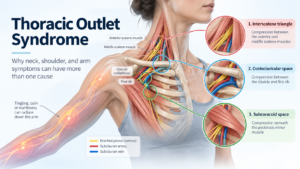
Our extended assessments can consider joint stability, biomechanics, fascial and myofascial tension, peripheral nerve irritation and vascular factors rather than looking at the painful area in isolation. When clinically appropriate, ultrasound can also support assessment and precision-guided procedures.
This broader approach matters because treating the tightest muscle is not always the same as treating the reason that muscle became tight.
Our goal is to understand the pattern, identify the likely pain generators and create a practical plan for what should happen next.
The bottom line
If you have hEDS or HSD and feel unusually tight, stiff or “restricted,” the sensation can seem contradictory.
Your joints may be highly mobile while your muscles are working extremely hard to control them.
Over time, this can contribute to muscle guarding, trigger points and myofascial pain.
Fascia may also contribute. It is a living, nerve-rich connective tissue that moves with our muscles and can participate in pain.
For selected patients, an ultrasound-guided fascial procedure such as hydrodissection may help reduce a local pain generator and improve comfortable movement.
But it is not a cure for hypermobility.
The most useful question is not simply:
“How do we release this tight tissue?”
It is:
“Why did this tissue become painful and protective in the first place—and what does the body need so it does not have to keep doing it?”
That is where a detailed assessment and an individualised treatment plan become important.
Concerned that hypermobility, trigger points or fascial pain may be contributing to your symptoms?
Laurel Clinical offers extended assessment for selected patients with hypermobility, EDS and complex musculoskeletal symptoms.
A detailed assessment can help clarify whether the problem is mainly related to joint instability, myofascial pain, nerve irritation, vascular factors or a combination of these—and whether an ultrasound-guided procedure has a useful role in your treatment plan.
This article provides general health information only and is not a substitute for individual medical assessment, diagnosis or treatment.
Sources and further reading
Hakim A. Hypermobile Ehlers-Danlos Syndrome. GeneReviews®, updated February 2024. This clinical reference describes joint instability, chronic soft-tissue problems, myofascial pain and current management principles for hEDS.
Suarez-Rodriguez V, Fede C, Pirri C, et al. Fascial Innervation: A Systematic Review of the Literature. International Journal of Molecular Sciences. 2022;23(10):5674. The review found substantial sensory innervation within fascia, including nociceptive and proprioceptive nerve endings.
Wu W-T, Chang K-V, Ricci V, Özçakar L. Ultrasound imaging and guidance in the management of myofascial pain syndrome: a narrative review. Journal of Yeungnam Medical Science. 2024;41(3):179–187.
Suarez-Ramos C, Gonzalez-Suarez C, Gomez IN, et al. Effectiveness of ultrasound guided interfascial hydrodissection with the use of saline anesthetic solution for myofascial pain syndrome of the upper trapezius: a single blind randomized controlled trial. 2023.
Chen Y, Liu S, Sun Y, et al. Efficacy of ultrasound-guided myofascial hydrodissection technique in myofascial pain syndrome of upper trapezius: a randomized controlled trial. Scientific Reports. 2025;15:33444.