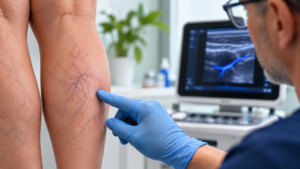


Patients sometimes come to see me with pain around the knee, convinced that their veins are responsible. They may have large varicose veins or small spider veins across the front of the knee. Naturally, they associate what they can see with what they can feel.
But when I ask them to show me exactly where the pain is, they often point somewhere else. The discomfort is deeper, sometimes behind the knee or towards its outer aspect.
It may still be related to venous disease. However, the location of the pain makes me consider other possibilities.
One structure I pay particular attention to is the popliteus muscle and its tendon. It sits deep in the back of the knee and is easily overlooked when the consultation begins with a concern about veins.
I encounter this situation in my work as a phlebologist. Ultrasound is central to my assessment, particularly because it allows me to investigate a suspected vascular problem and, when appropriate, examine some of the surrounding muscles and tendons.
The veins may be abnormal without causing all the pain
Varicose veins can certainly be symptomatic. Some patients experience swelling or aching, particularly after prolonged standing. Others have tender superficial veins.
When these findings correspond to the patient’s symptoms, the connection may be quite convincing.
Occasionally, though, the symptoms don’t fit.
A patient might have significant venous reflux on ultrasound but describe a very localised pain deep in the knee. The veins may explain the swelling, but I am less convinced that they account for the deeper discomfort.
I would be reluctant to recommend treating those veins with the expectation that every symptom will disappear.
The distinction is worth discussing with the patient because two conditions can be present at the same time. Treating one does not necessarily resolve the other.
What I look for on ultrasound
I usually begin by addressing the vascular concern that brought the patient to see me.
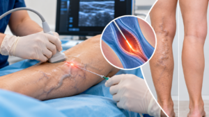
Venous duplex ultrasound allows me to assess the superficial and deep venous systems. I can look for reflux and investigate other venous abnormalities according to the clinical presentation. If deep vein thrombosis is suspected, it requires an appropriate and timely diagnostic assessment.
When the vascular findings don’t adequately explain the pain, I may extend my ultrasound assessment to the nearby musculoskeletal structures.
The popliteus is an interesting example.
It is a relatively small muscle involved in controlling rotation of the knee. Its tendon runs through the posterolateral part of the joint, where it can be examined using a targeted musculoskeletal ultrasound technique.
Published scanning techniques describe locating the tendon near the lateral femoral condyle and following its course through the popliteal sulcus.
The tendon is examined in both longitudinal and transverse planes. Careful adjustment of the transducer angle is necessary because normal tendon fibres can appear artificially dark when the ultrasound beam is not correctly aligned. This artefact, called anisotropy, can be mistaken for disease.
The examiner looks for changes in the tendon structure, including thickening or altered fibre appearance. Fluid around the tendon may also be relevant in some circumstances.
I am particularly interested in whether the ultrasound appearance corresponds to the patient’s symptoms.
For example, if the suspected tendon abnormality lies precisely where the patient describes pain, that finding deserves closer consideration. It becomes more convincing when the clinical examination supports the same explanation.
An abnormal image on its own is not enough.
There is also a limit to what ultrasound can establish. Deeper joint pathology may not be adequately assessed, and MRI can be useful when the diagnosis remains uncertain.
Sometimes the problem involves more than the tendon
Pain around the outer or back of the knee can also involve the peripheral nerves.
The common peroneal nerve travels around the fibular neck, close to the outer aspect of the knee. In selected patients, high-resolution ultrasound can help assess the nerve and surrounding tissues for a possible site of compression.
I consider nerve involvement particularly when the patient describes altered sensation or when the neurological examination suggests it.
Not every nerve problem will be apparent on ultrasound. Depending on the findings, further neurological assessment may be needed.
This broader assessment is useful when the original venous explanation seems incomplete.
A patient who had three contributing problems
I remember a patient with quite prominent varicose veins. They were causing discomfort and swelling, so there was a genuine venous problem to address.
However, the patient also experienced pain around the knee that I couldn’t satisfactorily attribute to the veins.
Further examination revealed pathology involving the popliteus tendon, along with entrapment of the peroneal nerve.
We discussed the findings and the different treatment options.
The patient wanted the varicose veins treated first. That was a reasonable preference given the symptomatic venous disease, and we proceeded accordingly.
The popliteus tendon problem was initially managed with physiotherapy. Unfortunately, the response was insufficient. Following reassessment, I subsequently performed ultrasound-guided injections as part of the management.
I mention this case because it illustrates something I frequently have to consider when assessing leg pain: the first abnormality we discover may be genuine without providing the complete diagnosis.
It also shows why the order of treatment is not necessarily dictated by the ultrasound findings alone. The patient’s preferences have a role, provided the proposed sequence is clinically appropriate.
In this case, identifying the additional musculoskeletal and neurological problems allowed us to address symptoms that could not be explained by venous disease alone.
Does finding popliteus tendinopathy mean an injection is needed?
Certainly not.
The ultrasound findings need to be considered alongside the severity of symptoms and their effect on the patient’s activities.
For some patients, appropriately directed rehabilitation may be sufficient. In others, further assessment may be necessary because the diagnosis remains uncertain or the response to treatment is disappointing.
I don’t consider an unsuccessful course of physiotherapy an automatic indication for an injection.
Before proceeding, I would want to reconsider whether the tendon really is the source of the pain. I would also review whether another condition could be contributing.
Ultrasound-guided injections have a role in selected musculoskeletal and nerve-related conditions, but the evidence varies considerably according to the specific pathology and treatment used.
They are not a routine solution for every painful tendon.
When an intervention is considered, the patient needs to understand its purpose and the uncertainty surrounding the expected benefit.
When should the investigation go further?
There are patients in whom the diagnosis becomes reasonably clear after clinical examination and a focused ultrasound assessment.
Others need additional investigation.
MRI may be appropriate when I suspect pathology involving deeper structures of the knee or when the ultrasound findings don’t satisfactorily explain persistent symptoms.
A sports physician or orthopaedic specialist may be better placed to assess certain mechanical knee problems. Patients with significant neurological findings may need dedicated nerve investigations.
I don’t see referral as a failure to reach a diagnosis. Sometimes it is the most appropriate recommendation.
A different situation arises when somebody develops sudden unexplained leg swelling or pain suggestive of deep vein thrombosis. That needs prompt medical assessment rather than being presumed to represent a tendon injury. Sudden breathlessness or chest pain warrants emergency assessment.
What I hope patients understand about their knee pain
People often come to a vein consultation expecting a fairly straightforward answer. They want to know whether their veins are responsible for the pain and whether treatment would help.
Sometimes I can give them that answer quite quickly.
Other times, the ultrasound shows a vascular abnormality that explains only part of what they are experiencing.
In those circumstances, I prefer to investigate the discrepancy rather than recommend a vein procedure on the assumption that it will solve everything.
The popliteus tendon is one possible explanation for pain behind the knee, but it is not the only one. Its assessment has to be guided by the clinical presentation.
For patients with both visible veins and unexplained knee pain, a consultation that considers the vascular findings alongside other possible sources of pain may provide a clearer direction.
At Laurel Clinical on the Gold Coast, I assess patients with venous disease and complex leg symptoms. When appropriate, ultrasound helps me explore whether the suspected vascular problem accounts for the symptoms or whether another explanation deserves attention.
The value of the consultation is not simply finding something abnormal on a scan. It is working out whether that finding is responsible for the patient’s symptoms and what, if anything, should be done about it.
Dr Gilles Laur is a Gold Coast doctor with qualifications including FACP, FRACGP, FASLMS and a Doctorate in Medicine. His clinical work includes phlebology, vascular ultrasound assessment and the evaluation of selected complex leg pain presentations.
This article is general medical information and does not replace an individual medical assessment.