
It sounds like a straightforward question. Someone has noticed fine red, blue or purple vessels on their legs. They may have become more prominent over time, or perhaps previous treatment has not produced the result they expected.
They want to know whether the veins can be removed, which treatment works best, and whether the results will last.
The short answer is that spider veins can often be significantly improved with treatments such as microsclerotherapy or vascular laser.
But in my practice, the more important question comes before choosing a treatment:
What are these visible veins telling me about the venous system underneath?
Most spider veins are not a sign of serious disease. However, assuming that every spider vein is simply a cosmetic problem can sometimes mean overlooking important information.
My approach is to understand the pattern before deciding what, if anything, should be treated.
Spider veins are visible. Their underlying anatomy is not always obvious.
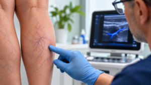
When I examine spider veins, I pay attention to more than their colour or size.
I study their shape, distribution and organisation. Are they scattered individually, arranged in clusters, or connected to larger blue-green reticular veins beneath the skin?
Are they concentrated in a particular anatomical area? Is there a pattern suggesting an underlying feeding vein or a connection with a perforating vein?
These details influence how I interpret what I see.
I also want to know what the patient experiences.
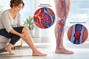
Do their legs feel heavy or painful? Do they swell towards the end of the day? Have they previously undergone venous treatment? Is there a history of deep vein thrombosis?
I look for skin changes such as brown pigmentation, venous eczema or ulceration, which can suggest more advanced venous disease.
None of these findings should be interpreted in isolation.
For example, a patient may have extensive spider veins without significant underlying venous disease. Another may have only a small visible cluster but also swelling, skin changes or a history suggesting a more complicated venous problem.
The number of spider veins someone has does not necessarily reflect the significance of their underlying venous condition.
Equally, leg pain should not automatically be attributed to spider veins merely because the two occur together.
The task is to determine what is related, what may be incidental, and what deserves further assessment.
Why do I use ultrasound when assessing spider veins?
One reason I approach spider veins carefully is that experience has taught me not to underestimate an apparently simple presentation.
I have encountered a situation in which spider veins that initially appeared straightforward were associated with an unusual venous malformation extending into the region of the sciatic nerve.
This is not a common explanation for spider veins, and I would never suggest that most people have a hidden vascular malformation.
However, it reinforced an important principle in my clinical practice: the visible appearance does not always reveal the underlying anatomy.
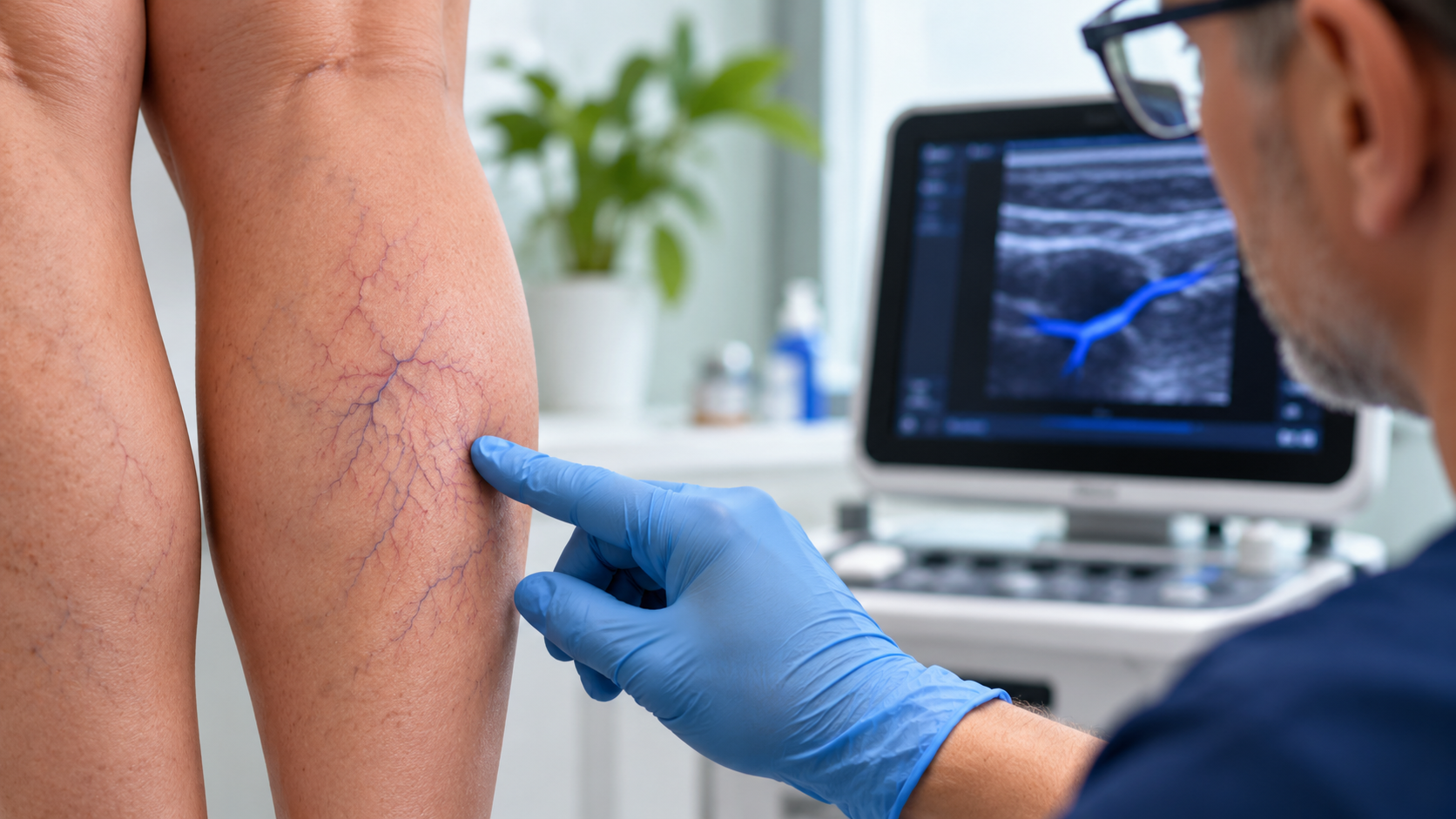
I routinely incorporate venous duplex ultrasound into my assessment of patients presenting for leg spider-vein treatment.
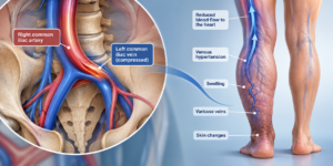
My aim is to evaluate the broader venous system and identify relevant abnormalities, including reflux, associated reticular networks, perforating veins and other findings that could influence management.
Ultrasound does not visualise every tiny surface vessel, but it can help identify larger anatomical connections and assess blood flow.
There is an important distinction here.
Clinical guidelines do not recommend routine duplex ultrasound for every person with isolated, symptom-free spider veins. My use of ultrasound reflects my own assessment approach, informed partly by the unusual conditions I have encountered.
Finding an abnormality on ultrasound does not automatically mean that the abnormality is causing the spider veins, or that it should be treated.
An investigation is valuable when its findings can meaningfully influence a decision. It should not become a justification for an unnecessary procedure.
Is sclerotherapy or laser better for spider veins?
Patients often ask which technology produces the best results.
I do not believe one treatment should automatically be preferred for every spider vein.
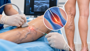
Microsclerotherapy involves injecting a sclerosant into selected small veins, producing a controlled reaction that can lead to their closure and gradual disappearance.
Vascular laser delivers targeted energy to selected vessels, producing a different biological response.
Both techniques have advantages, limitations and potential complications.
The choice depends on factors including vessel diameter, depth, distribution, associated feeding veins, patient characteristics and the findings of the clinical assessment.
Some vessels are better suited to injection treatment. Others may be more appropriately treated with a vascular laser. Some presentations may benefit from combining techniques.
Sometimes the important decision is not which technique to use, but which veins should be addressed first.
If an underlying venous network is contributing to the visible vessels, that connection may influence treatment sequencing. However, not every abnormal vein detected on ultrasound requires intervention.
I do not select a treatment simply because the equipment is available. I select it because its characteristics, benefits and risks make sense for the particular vein and patient.
The technology matters, but so do interpretation, treatment planning and technique.
When would I recommend against treating spider veins?
Not every spider vein needs treatment, even when treatment is technically possible.
If someone has a few very fine vessels causing little concern, observation may be entirely reasonable.
Some vessels are too small to be treated reliably with the available techniques. Attempting to eliminate every microscopic vessel may increase treatment burden without producing a meaningful improvement.
I am also cautious when there are other problems that deserve attention first.
For example, if a patient has impaired calf-muscle pump function, swelling or another significant venous disorder, I may need to address those issues before considering cosmetic treatment.
Contraindications to sclerotherapy or laser treatment, previous complications, relevant medical conditions and individual risk factors can also change my recommendation.
But one of the most important considerations is the patient’s expectation.
If someone expects completely flawless legs, with no visible vessels and no possibility of recurrence, I would be concerned that treatment may not meet their expectations.
I explain that even appropriately performed procedures can cause complications.
These may include bruising, temporary discomfort, pigmentation changes, new fine vessels known as telangiectatic matting, or unwanted skin changes. More serious complications, including tissue injury, allergic reactions or venous thrombosis, are uncommon but must be considered. Risks differ between treatments.
No clinician can eliminate every risk.
When treatment is elective, the patient must consider whether the likely improvement is worth the possible complications. Choosing not to proceed is a legitimate medical decision.
Sometimes the appropriate recommendation is to do nothing for now.
Can spider veins disappear completely and permanently?
Many spider veins can fade considerably after treatment, and some may disappear from view.
However, complete and permanent clearance cannot be promised.
When I discuss expected results, I distinguish between improvement in the vessels we can treat and the long-term tendency to develop additional veins.
A treated vessel may close successfully, but other small vessels can become visible over time.
Some may already be present but too fine to treat reliably. Others may develop or enlarge later as the underlying factors affecting the venous system continue.
This is why I do not regard spider-vein treatment as a single, guaranteed endpoint.
The response also varies between individuals.
Treatment causes biological changes in the vessel and surrounding tissues. People do not all respond identically.
Some patients achieve substantial improvement with relatively limited treatment. Others require several sessions, may have incomplete clearance, or develop pigmentation or matting.
It is important to discuss these possibilities before treatment rather than explaining them only after an unexpected result.
I also consider ways to support venous function over time.
Calf-muscle strengthening and appropriately selected compression stockings may be useful in managing certain venous symptoms or functional problems. However, they should not be presented as proven methods of preventing all future spider veins.
A successful treatment is not necessarily one that removes every visible vessel. It is one that achieves a worthwhile improvement at a level of risk the patient understands and accepts.
What if my spider veins keep coming back?
When spider veins reappear after treatment, I want to understand what has actually happened.
Are these previously treated vessels that did not fully respond? Have new vessels developed? Are there associated reticular veins or another venous problem that may be contributing?
The distinction matters because repeating the same treatment without reconsidering the underlying pattern may not be the most useful approach.
Sometimes additional treatment is reasonable. Sometimes a different technique or sequence is appropriate. Occasionally, the more important decision is to investigate or manage another condition.
A recurrence does not automatically mean that previous treatment was performed incorrectly. Nor does it mean the patient necessarily needs a more aggressive procedure.
It means the situation deserves reassessment.
Who may particularly benefit from a more detailed venous assessment?
My work in phlebology on the Gold Coast includes patients with spider veins, varicose veins, swollen legs, venous malformations, post-thrombotic conditions and more complex venous presentations.
A detailed opinion may be particularly useful when spider veins occur alongside persistent swelling, leg heaviness, pain, skin changes, previous thrombosis or recurring problems after treatment.
These situations can involve questions beyond the appearance of the superficial veins.
However, someone with a small number of uncomplicated spider veins and no associated symptoms may have much simpler needs.
The level of investigation and treatment should remain proportionate to the clinical question and the patient’s goals.
My final perspective: Treat the patient, not just the visible veins
The question “Can I get rid of my spider veins?” usually has an encouraging answer: meaningful improvement is often possible.
But whether treatment is appropriate, how it should be performed, and what degree of improvement is realistic are separate questions.
My experience has taught me to respect apparently simple cases without assuming that they are all complicated.
I want to understand what the visible veins represent, whether an underlying finding is clinically important, which treatment offers a reasonable balance of benefit and risk, and whether the patient would genuinely benefit from proceeding.
The value of a clinical opinion is not simply knowing how to remove a vein. It is knowing which findings matter, which treatment is justified, and when leaving something alone may be the better choice.
If your spider veins are associated with swelling, pain, skin changes or previous unsuccessful treatment, an individual venous assessment may help clarify the options.
And if your concern is purely cosmetic, an informed discussion about realistic results and treatment risks can be just as valuable.
Dr Gilles Laur is a Gold Coast medical practitioner whose clinical work includes phlebology, venous ultrasound assessment and the management of superficial and complex venous conditions.
This article provides general medical information and does not replace an individual assessment. Treatment suitability and results vary. New or sudden one-sided leg swelling or pain requires prompt medical assessment rather than cosmetic vein treatment.