
If you have varicose veins, a proper assessment should do more than look at the veins you can see.
The goal is to understand why the veins have developed, where abnormal blood flow is coming from, whether deeper veins are involved, and whether your symptoms are actually being caused by vein disease.
What should a comprehensive varicose vein assessment include?
A comprehensive varicose vein assessment should include six main parts:
- Medical and previous-treatment history
- Assessment of symptoms and how they affect daily life
- A clinical examination, usually with the patient standing
- Duplex ultrasound mapping of the superficial and deep veins
- Identification of the true source and pathway of reflux or obstruction
- Assessment for less common or atypical causes when the findings do not explain the symptoms
Duplex ultrasound is an important part of this process. Guidelines recommend it for assessing symptomatic varicose veins because it can show the pattern of abnormal blood flow and help plan treatment.
Here is what each part of the assessment means and why it matters.
1. Medical and Previous-Treatment History
A good varicose vein assessment starts with a conversation.
Your clinician needs to understand what has happened before looking at what is happening now.
You may be asked about:
- When your varicose veins first appeared
- Whether they have become worse
- Previous pregnancies
- A family history of vein problems
- Previous blood clots, including deep vein thrombosis (DVT)
- Previous leg injuries or surgery
- Other health conditions
- Medicines you take
- Previous varicose vein treatments
- Whether treated veins have returned
Previous treatment is especially important.
If you have already had laser treatment, radiofrequency treatment, sclerotherapy or vein surgery, new varicose veins may have developed through a different pathway. The clinician needs to know what was treated before deciding what should happen next.
A history of DVT is also important because a previous clot can damage deeper veins and affect how blood leaves the leg.
2. Symptoms and Their Impact on Daily Life
Varicose veins do not affect everyone in the same way.
Some people have large visible veins with few symptoms. Others may have aching, heaviness or swelling that makes it difficult to work, exercise or stand for long periods.
Common symptoms linked with venous disease can include:
- Aching
- Pain or discomfort
- Heavy legs
- Swelling
- Itching
- Throbbing
- Tired or restless legs
- Symptoms that become worse after standing
- Skin irritation or changes around the ankle
NICE identifies pain, aching, discomfort, swelling, heaviness and itching among the typical symptoms associated with symptomatic varicose veins.
But simply having leg pain does not prove that varicose veins are the cause.
That is why a good assessment asks when symptoms occur, what makes them better or worse, and how much they affect everyday activities.
For example, does the leg become heavy after standing at work? Does swelling increase toward the end of the day? Does discomfort improve after walking or raising the leg?
These details help the clinician decide whether the symptoms fit a venous pattern.
3. A Standing Clinical Examination
Varicose veins can look very different when you are lying down compared with when you are standing.
When you stand, gravity increases the pressure within the veins of the legs. This can make abnormal veins and swelling easier to see.
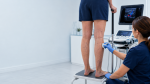
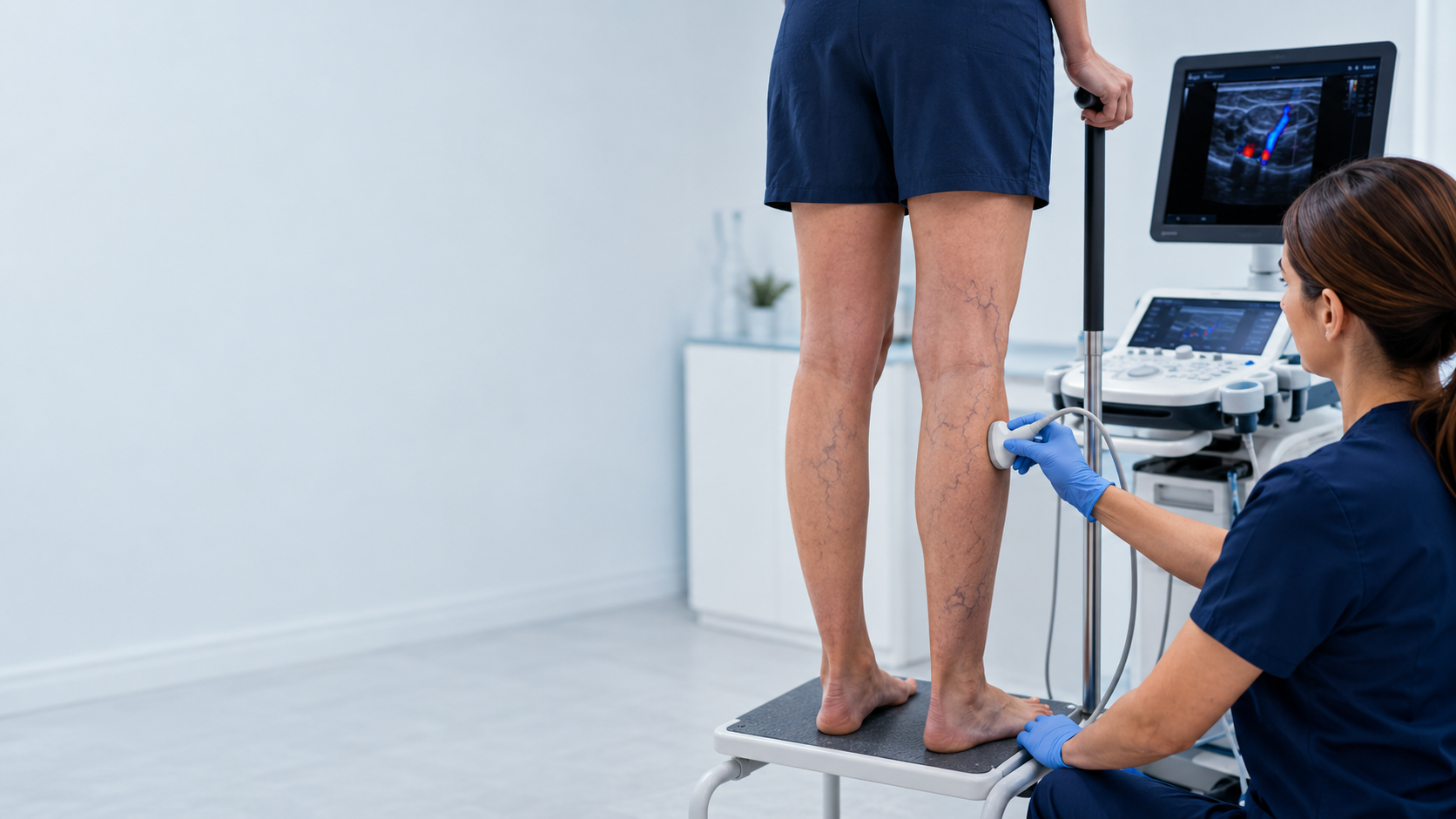
For this reason, examination of varicose veins commonly includes looking at the legs while the patient is standing.
The clinician may examine both legs for:
- Visible varicose veins
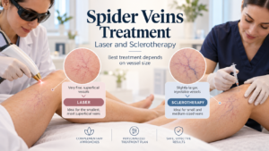
- Spider or reticular veins
- Ankle or lower-leg swelling
- Skin discolouration
- Venous eczema
- Hard or inflamed veins
- Changes in the skin or tissue
- Previous scars
- Healed or active ulcers
- Unusual patterns of veins
The location of the veins can provide useful clues.
For example, veins around the inner thigh may have a different source from veins behind the calf. Unusual veins around the groin, abdomen or pelvis can sometimes suggest that the problem is coming from higher up.
The clinician may also check the circulation in the arteries of the legs, particularly if there are symptoms or signs suggesting that arterial disease could also be present.
4. Duplex Ultrasound Mapping of the Superficial and Deep Veins
For people with symptomatic varicose veins, looking at the leg is only part of the assessment.
The veins underneath the skin also need to be examined.
This is usually done with a duplex ultrasound scan.
Duplex ultrasound combines an image of the veins with information about the direction and speed of blood flow. It can show whether veins are open and whether their valves are working correctly.
NICE recommends duplex ultrasound to confirm varicose veins, determine the extent of reflux and help plan treatment.
What should the ultrasound examine?
A comprehensive scan may assess both the superficial veins and the deep veins.
The superficial system includes veins close to the skin, such as the great and small saphenous veins and their branches.
The deep system contains the larger veins that carry most of the blood back toward the heart.
This distinction matters because visible varicose veins can sometimes be connected with problems elsewhere in the venous system.
Current vascular guidelines recommend evaluating the deep venous system in patients with symptomatic varicose veins for problems such as obstruction or faulty valves.
Should a vein ultrasound be performed standing?
For reflux assessment, the veins should normally be examined with gravity acting on them.
The Society for Vascular Surgery, American Venous Forum and American Vein and Lymphatic Society recommend performing reflux ultrasound with the patient standing whenever possible. Alternative positions can be used when a patient cannot safely stand.
This helps the clinician see how the veins behave under conditions closer to normal everyday life.
5. Identifying the Actual Source and Pathway of Reflux or Obstruction
This is one of the most important parts of a comprehensive varicose vein assessment.
It is not enough to find a varicose vein.
The clinician should try to determine where the abnormal blood flow starts and where it travels.
What is venous reflux?
Healthy leg veins contain valves that help blood travel upward toward the heart.
If these valves do not close properly, some blood can move backwards. This backward flow is called venous reflux.
Over time, the extra pressure can contribute to enlarged and twisted superficial veins.
But reflux can occur in different places.
It may involve:
- A major superficial vein
- A junction between veins
- A smaller branch
- A perforating vein connecting superficial and deep veins
- The deep venous system
- In selected cases, veins originating from the pelvis
Current specialist guidelines state that superficial reflux should be traced back to its source rather than simply recording that reflux is present.
This is important when planning treatment.
Think of a varicose vein like water appearing downstream from a leaking pipe. Treating only the part you can see may not solve the problem if the main leak is somewhere else.
What about venous obstruction?
Not every vein problem is caused by reflux.
Sometimes blood has difficulty travelling out of the leg because a vein is narrowed or blocked. This is known as venous obstruction.
For example, previous DVT can sometimes leave scarring or narrowing within the deep veins.
Symptoms such as significant swelling, leg or thigh fullness, heaviness, or discomfort with walking may lead the clinician to investigate whether a deeper obstruction is present.
Guidelines recommend further assessment for iliofemoral obstruction in patients with signs or symptoms that suggest a problem higher in the venous system.
6. Looking for Atypical Causes When the Findings Do Not Explain the Symptoms
Sometimes the examination and ultrasound findings do not fully explain what a person is experiencing.
That should not automatically mean that more visible veins need to be treated.
Instead, the clinician may need to ask a different question:
Could something else be causing these symptoms or this unusual vein pattern?
Possible issues that may need consideration include:
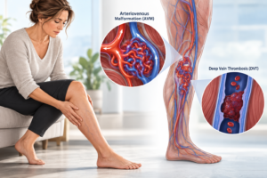
- Previous deep vein thrombosis
- Deep venous obstruction
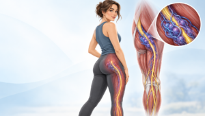
- Pelvic sources of varicose veins
- Unusual vein anatomy
- Arteriovenous malformations
- Lymphatic swelling
- Arterial circulation problems
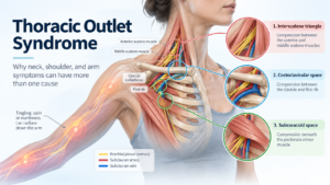
- Musculoskeletal causes of leg pain
- Nerve-related symptoms
Which conditions need investigation will depend on the person’s history, examination and ultrasound findings.
Specialist venous guidelines specifically recommend further investigation for deeper or pelvic venous problems when certain symptoms or unusual patterns of veins are present.
This part of the assessment is important because not every painful or swollen leg is caused by superficial varicose veins.
Why Is a Full Varicose Vein Assessment Important Before Treatment?
Modern varicose vein treatment can be highly targeted.
But targeted treatment requires an accurate diagnosis first.
A proper assessment should answer questions such as:
- Are the visible veins actually causing the symptoms?
- Which veins have abnormal reflux?
- Where does that reflux begin?
- Are the deep veins working normally?
- Is there any evidence of obstruction?
- Have previous treatments changed the vein anatomy?
- Is there another possible cause for the symptoms?
- Which treatment, if any, best matches the problem?
This is why an ultrasound should not simply be used to confirm that a varicose vein exists.
It should help create a map of the venous problem.
NICE describes a specialist vascular service as one capable of carrying out a full clinical and duplex ultrasound assessment and providing appropriate treatment options.
Is an Ultrasound Always Needed for Varicose Veins?
Not every tiny visible vein automatically requires a detailed ultrasound.
For example, international guidelines do not recommend routine duplex scanning for people who have only small, asymptomatic spider or reticular veins, because unnecessary scanning can sometimes lead to unnecessary treatment.
The situation is different when varicose veins are causing symptoms, have returned after previous treatment, or are associated with complications.
In these cases, duplex ultrasound can provide important information that cannot be obtained by simply looking at the leg.
When Should Varicose Veins Be Assessed?
Consider seeking medical assessment if varicose veins are associated with:
- Persistent aching or pain
- Leg heaviness
- Swelling
- Itching or eczema
- Skin discolouration
- Hard or painful veins
- A previous or current leg ulcer
- Recurrent varicose veins after treatment
Bleeding from a varicose vein requires prompt medical attention. NICE recommends immediate referral to a vascular service for bleeding varicose veins.
Sudden one-sided leg swelling or pain can also have causes other than ordinary varicose veins, including DVT, and should be medically assessed.
The Bottom Line
A comprehensive varicose vein assessment is not simply an examination of the veins that are visible through the skin.
It should combine your medical history, your symptoms, a standing clinical examination and detailed duplex ultrasound mapping.
Most importantly, the assessment should identify the actual source and pathway of abnormal blood flow, including reflux or obstruction.
If those findings do not explain the symptoms, a good assessment should consider whether another condition or a less common venous problem could be responsible.
Finding the source of the problem first allows treatment decisions to be based on the underlying vein anatomy and blood flow rather than appearance alone.
Frequently Asked Questions
What is included in a varicose vein assessment?
A comprehensive assessment normally includes your medical history, symptoms, previous vein treatments, examination of the legs, duplex ultrasound of relevant superficial and deep veins, and identification of the source of abnormal reflux or obstruction.
What is the best test for varicose veins?
Duplex ultrasound is the main diagnostic imaging test used to investigate symptomatic varicose veins. It shows the anatomy of the veins as well as the direction of blood flow.
Why do doctors examine varicose veins while standing?
Standing allows gravity to fill the veins and can make varicose veins and abnormal reflux easier to assess. Clinical examination and venous reflux ultrasound are therefore commonly performed with the patient standing when possible.
Can varicose veins come back after treatment?
Yes. New or recurrent varicose veins can occur after previous treatment. This is one reason a new assessment should include a history of earlier procedures and an ultrasound map of the current venous circulation.
Can leg pain be caused by something other than varicose veins?
Yes. Leg pain, swelling and heaviness can have many causes. If the pattern of varicose veins or ultrasound findings does not explain the symptoms, further assessment may be needed to look for venous obstruction or non-venous causes.
Why is finding the source of reflux important?
The visible varicose vein may be only one part of a larger pathway of abnormal blood flow. Finding where reflux begins helps the clinician understand which vein is causing the problem and plan treatment appropriately.
This article provides general health information and is not a substitute for individual medical advice, diagnosis or treatment.