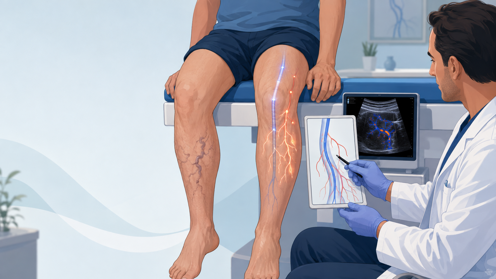

If you have had EVLA treatment and your varicose veins have come back, you may wonder: Did the laser treatment fail?
Not necessarily.
Varicose veins can recur after EVLA for several different reasons. Sometimes the vein that was treated opens again. But in many people, the original vein stays successfully closed and a different vein develops a problem later.
The important question is not simply, “Have the varicose veins come back?”
It is:
Where is the abnormal blood flow coming from now?
A repeat duplex ultrasound can usually help answer that question.
What is EVLA?
EVLA stands for endovenous laser ablation.
It is a minimally invasive treatment used to close a vein that has faulty valves and allows blood to flow backwards. This backward flow is called venous reflux.
EVLA is commonly used to treat reflux in the great saphenous vein or small saphenous vein.
A thin laser fibre is placed inside the abnormal vein. Laser energy heats the vein wall, causing the vein to close. Blood then travels through healthier veins instead.
EVLA can successfully treat the problem that exists at the time of treatment. However, it cannot stop every other vein in the leg from developing valve problems in the future.
That is one reason varicose veins can recur.
Why do varicose veins come back after EVLA?
There are several possible causes of recurrent varicose veins after EVLA.
Some are related to the vein that was originally treated. Others happen because venous disease has developed somewhere else.
1. The treated vein can reopen
One possible cause is recanalisation.
This means that a vein that was previously closed by EVLA develops an open channel again and blood begins flowing through it.
The whole vein does not always reopen. Sometimes only a small section does.
More importantly, seeing some blood flow in the vein does not automatically mean it is causing a problem. The duplex ultrasound needs to show whether there is significant backward flow, or reflux, and whether that reflux is feeding the new varicose veins.
If the previously treated vein has reopened and developed significant reflux, this can be a true cause of recurrence.
2. Another saphenous vein can become faulty
The leg contains more than one superficial vein.
A patient may have successful EVLA of the great saphenous vein but later develop reflux in an accessory saphenous vein.
One important example is the anterior saphenous vein, which runs along the front and side of the thigh.
This vein may have been completely normal when the original treatment was performed.
Years later, its valves may stop working properly. Blood can then flow backwards through it and produce a new network of varicose veins.
In this situation, the original EVLA has not necessarily failed.
The vein that was treated may still be completely closed. A different vein has become incompetent.
3. Some smaller veins may remain after EVLA
EVLA mainly treats the larger vein responsible for the abnormal reflux.
The visible, twisted veins under the skin are often branches called tributaries.
After EVLA, some of these smaller veins shrink by themselves because the pressure feeding them has been removed.
Others do not disappear completely.
These are sometimes called residual tributaries.
They may remain visible after treatment or become larger again if they later connect with another source of reflux.
Depending on the individual case, these veins may sometimes require additional treatment such as phlebectomy or ultrasound-guided sclerotherapy.
4. Perforator veins can become incompetent
Perforator veins connect veins near the surface of the leg with the deeper veins.
They normally help direct blood towards the deep venous system.
Sometimes the valves inside a perforator vein stop working properly. Blood can then move in the wrong direction and increase pressure in nearby surface veins.
An incompetent perforator may contribute to recurrent varicose veins, particularly when the new veins are concentrated in one area of the thigh or calf.
However, finding a perforator on ultrasound does not automatically mean it is the cause.
The sonographer needs to determine whether abnormal flow from that perforator actually connects with and feeds the recurrent veins.
5. Venous disease can progress over time
This is one of the most important points to understand.
Varicose veins are usually part of an ongoing condition called chronic venous disease.
EVLA treats the abnormal veins that are present at that particular time. It does not permanently protect every vein in the leg from developing valve problems later.
Imagine repairing one leaking pipe in a plumbing system. The repair may remain perfect, but another pipe could develop a problem several years later.
Something similar can happen with veins.
A patient could have successful EVLA, feel much better for five or ten years and then develop new varicose veins from a completely different vein.
That does not mean the original EVLA suddenly stopped working.
It may mean the underlying venous disease has progressed.
6. The reflux can sometimes come from the pelvis
Not every varicose vein in the leg starts inside the leg.
In some patients, abnormal blood flow can travel from veins in the pelvis into veins around the groin and upper leg. This is called pelvic venous reflux.
Pelvic reflux may be considered when unusual varicose veins appear around the groin, vulval or perineal area, inner thigh, back of the thigh or buttock.
This does not mean everyone with recurrent varicose veins needs a pelvic scan.
Most do not.
But if the pattern of veins suggests that blood is entering the leg from the pelvis, further investigation may be appropriate.
7. There may occasionally be a blockage higher up
In selected patients, recurrent venous problems may be related to difficulty getting blood out of the leg.
The larger veins inside the pelvis are called the iliac veins.
If one of these veins is significantly narrowed or obstructed, pressure can build up below the obstruction. The body may create alternative pathways for blood to return towards the heart.
These pathways can sometimes appear as unusual or recurrent varicose veins.
Previous deep vein thrombosis is one possible cause of this type of problem.
Iliac vein obstruction is not something that needs to be investigated in every person with recurrent varicose veins. It becomes more relevant when the patient’s symptoms, examination or duplex ultrasound suggest a problem higher up.
Does recurrent varicose disease mean EVLA failed?
No. Recurrent varicose veins do not automatically mean EVLA failed.
There is an important difference between the treated vein reopening and new disease developing elsewhere.
For example, imagine a patient had EVLA of the great saphenous vein five years ago.
A new duplex scan shows that the treated great saphenous vein is still completely closed. However, an anterior saphenous vein has now developed significant reflux and is feeding new varicose veins.
The patient has recurrent varicose veins.
But the original EVLA is still working.
That is why recurrence should not be diagnosed simply by looking at the veins on the skin.
The source of the abnormal blood flow needs to be identified.
What should a repeat duplex ultrasound check?
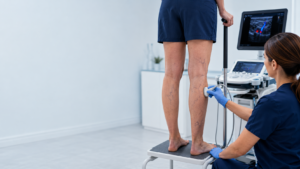
A duplex ultrasound is one of the most important tests for recurrent varicose veins because it can show both the anatomy of the veins and the direction of blood flow.
A good repeat duplex should not simply answer, “Is the old vein still closed?”
It should map the venous system and find the new source of reflux. Depending on the patient’s history and symptoms, the examination may need to assess:
the entire vein that was previously treated, looking for recanalisation and reflux;
the saphenofemoral junction in the groin or saphenopopliteal junction behind the knee;
the great and small saphenous veins, including untreated sections;
accessory saphenous veins, especially the anterior saphenous vein;
the visible recurrent varicose veins and what is feeding them;
important perforator veins;
the deep veins for reflux, previous thrombosis or signs of obstruction;
the common femoral vein in the groin for signs that could suggest a problem higher in the pelvis; and
possible pelvic sources when the pattern of recurrent veins suggests reflux is entering the leg from above.
The aim is to trace the reflux back to its source.
That information helps determine what treatment, if any, is appropriate.
When should recurrent varicose veins be investigated above the groin?
Most people with recurrent varicose veins do not automatically need pelvic or abdominal investigations.
However, investigation above the groin may be considered when the clinical examination or duplex scan suggests that the problem could be coming from the pelvis or from obstruction in the iliac veins.
Warning signs can include unusual veins around the groin or pelvic region, veins crossing the lower abdomen or pubic area, significant swelling mainly affecting one leg, a history of a major deep vein thrombosis, symptoms suggesting difficulty draining blood from the leg, or abnormal blood-flow patterns in the common femoral vein on duplex ultrasound.
The decision should be based on the whole clinical picture rather than on the presence of recurrent varicose veins alone.
Can recurrent varicose veins be treated again?
Often, yes.
But the correct treatment depends on why the veins have returned.
If the original vein has reopened, one approach may be appropriate.
If a new accessory vein is causing the reflux, treatment needs to target that vein instead.
If the problem is a residual tributary, perforator, pelvic source or obstruction higher in the venous system, the treatment strategy may be different again.
This is why repeating treatment before properly mapping the veins can be a mistake.
The first step should be finding the source.
Frequently Asked Questions
Why have my varicose veins returned after laser treatment?
They may have returned because the treated vein reopened, another superficial vein developed reflux, residual branches remained, perforator veins became incompetent or chronic venous disease progressed. Less commonly, reflux may originate from pelvic veins or there may be obstruction higher in the venous system.
Can a vein reopen after EVLA?
Yes. This is called recanalisation. It may involve part or all of the treated vein. Duplex ultrasound can determine whether the reopened section has significant reflux.
Can new varicose veins develop even if EVLA worked?
Yes. The original treated vein can remain successfully closed while another vein becomes incompetent later. This is one reason recurrent veins do not automatically mean EVLA failed.
How do you find the cause of recurrent varicose veins?
A detailed duplex ultrasound is usually the starting point. It should map the recurrent veins and trace abnormal blood flow back to its source rather than simply checking the previously treated vein.
Do recurrent varicose veins mean I need another operation?
No. Some recurrent veins do not require treatment, while others can be managed using minimally invasive procedures. The appropriate treatment depends on symptoms, anatomy and the source of reflux.
When do recurrent varicose veins need pelvic investigation?
Pelvic or abdominal investigation may be considered when the location of the recurrent veins, symptoms or duplex findings suggest pelvic venous reflux or obstruction above the groin. It is not routinely necessary for every patient.
The bottom line
Varicose veins can recur after EVLA without the original treatment having failed.
The treated vein may occasionally reopen, but recurrence can also happen because another saphenous vein becomes incompetent, residual tributaries remain, perforator veins develop reflux or chronic venous disease progresses.
In selected patients, the source may even be above the groin because of pelvic venous reflux or problems with venous outflow.
So when varicose veins return, the important question is not simply:
“Have my veins come back?”
It is:
“Where is the reflux coming from now?”
A properly performed repeat duplex ultrasound can usually provide the answer and guide the most appropriate next step.