
How Are Vascular Compression Syndromes Diagnosed? Why One Scan Is Not Enough
An evidence-based guide to symptoms, Doppler ultrasound, CT and MRI, specialist review and selective invasive testing — updated August 2026
Vascular compression syndromes can be confusing.
A scan may show that a vein or artery is being squeezed by another structure. It is tempting to assume that this finding explains a person’s symptoms.
But medicine is not usually that simple.
Finding compression on a scan is not always the same as diagnosing a vascular compression syndrome.
Some people have quite noticeable compression on imaging but have no symptoms at all. This has been shown with conditions such as iliac vein compression and left renal vein compression. Modern reviews therefore stress that diagnosis should combine the clinical picture with the right imaging findings.
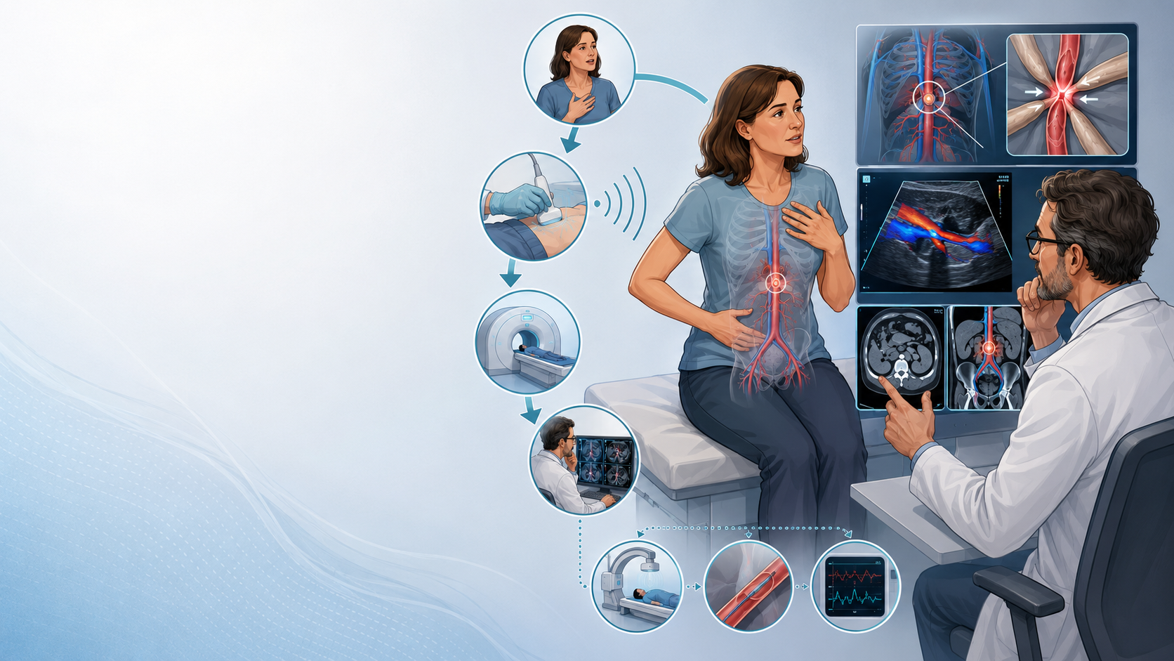
The safest way to think about diagnosis is as a hierarchy:
1. Does the person have the right clinical pattern?
2. Can targeted Doppler ultrasound show an abnormal effect on blood flow?
3. Does CT or MRI show the anatomy and support the suspected diagnosis?
4. Do the symptoms and imaging findings make sense together when reviewed by an experienced specialist?
5. Is a more invasive test actually needed before making a treatment decision?
That process is much more reliable than treating one measurement on one scan as a diagnosis.
First: what is a vascular compression syndrome?
A vascular compression happens when an artery or vein is pressed on by another structure in the body.
Depending on where this happens, the compressed vessel may carry less blood, develop abnormal pressure, or cause blood to be redirected through other veins.
Examples include:
Nutcracker syndrome, involving compression of the left renal vein;
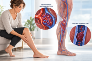
iliac vein compression, sometimes called May–Thurner syndrome when it causes a compatible clinical problem;
median arcuate ligament syndrome (MALS), involving compression around the celiac artery;
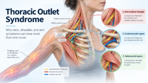
and vascular thoracic outlet syndrome, involving the subclavian artery or vein near the shoulder and upper chest.
These conditions affect different blood vessels and produce different symptoms, so their exact diagnostic tests are not identical.
But they share one very important rule:
An abnormal-looking blood vessel does not automatically mean the person has a syndrome.
The finding has to make sense with the person’s symptoms and the way blood is actually flowing.
Anatomy is not the same as disease
This is one of the most important ideas in vascular compression medicine.
Take iliac vein compression.
The left common iliac vein can be squeezed between an artery and the spine. This anatomy can be seen in people who have symptoms and in people who feel completely well. A 2024 multidisciplinary consensus statement specifically notes that iliac vein compression occurs in both symptomatic and asymptomatic people.
The same problem occurs with nutcracker anatomy.
Nutcracker phenomenon generally describes compression of the left renal vein seen on imaging. Nutcracker syndrome means that the compression is linked to a compatible clinical problem. A 2025 international Delphi consensus highlighted that asymptomatic renal-vein compression is common enough that imaging has to be interpreted alongside the clinical picture. Experts also failed to agree on one imaging cut-off that can diagnose the syndrome by itself.
A similar issue exists with celiac artery compression in MALS. Modern MALS literature recognises asymptomatic celiac compression and continues to describe the diagnosis as challenging because symptoms can overlap with many other conditions.
So the first question should usually not be:
“Is there compression?”
It should be:
“Is there evidence that this compression is causing the person’s clinical problem?”
Step 1: Start with the clinical phenotype
The clinical phenotype simply means the pattern of symptoms, signs and medical history.
This should come before trying to diagnose a compression from a scan.
Different compression syndromes tend to produce different clinical patterns.
Nutcracker syndrome
Nutcracker syndrome can cause symptoms such as:
blood in the urine;
left-sided abdominal or flank pain;
protein in the urine, especially related to posture;
varicocele in males;
and some forms of pelvic or gonadal venous congestion.
A 2026 review stresses that diagnosis requires careful clinical assessment, exclusion of other possible causes and targeted imaging.
Iliac vein compression
Symptomatic iliac vein compression may be considered in people with problems such as:
unexplained one-sided leg swelling;
venous claudication, where the leg becomes painful or heavy with activity;
chronic venous symptoms;
an iliofemoral deep vein thrombosis;
or certain patterns of pelvic venous disease.
The American College of Radiology’s 2026 guidance emphasises that iliac vein compression is often clinically silent. A careful history and examination are therefore needed to look for other causes before imaging findings are labelled as a syndrome.
Median arcuate ligament syndrome
MALS usually enters the differential diagnosis when a patient has a compatible pattern such as ongoing upper abdominal pain, often after meals, with symptoms that may include nausea, vomiting and weight loss.
These symptoms are not specific to MALS. They can occur in many gastrointestinal conditions. Recent literature therefore continues to stress careful clinical assessment and investigation for other possible explanations.
Vascular thoracic outlet syndrome
Vascular thoracic outlet syndrome affects the blood vessels passing between the upper chest and arm.
Venous compression may cause arm swelling, heaviness or discolouration and can be linked with thrombosis of the axillary or subclavian vein.
Arterial compression may produce arm or hand ischaemic symptoms and can sometimes cause arterial injury or embolisation.
Current reviews emphasise that the patient’s history and physical examination remain central, with imaging then used to identify which structure is being compressed and whether the finding is clinically important.
Step 2: Use targeted Doppler duplex ultrasound
Once the clinical picture raises a reasonable suspicion, Doppler duplex ultrasound is often useful.
Duplex ultrasound combines ordinary ultrasound pictures with Doppler measurements of blood flow.
That means it can sometimes answer two questions:
What does the blood vessel look like?
and
What is happening to blood flow through it?
This is valuable because vascular compression syndromes are not only about anatomy. They are also about whether the compression has an important effect on circulation.
But the ultrasound needs to be targeted to the suspected condition.
A routine leg-vein ultrasound looking for a blood clot is not necessarily the same as an examination designed to investigate iliac vein compression.
Likewise, an ultrasound for suspected nutcracker syndrome needs to examine the left renal vein and its blood-flow pattern.
For MALS, Doppler assessment of the celiac artery may include measurements during different phases of breathing because compression can become more pronounced during expiration.
For vascular thoracic outlet syndrome, a specialised ultrasound may assess the vessels with the arm in different positions because the compression can be dynamic. A 2026 review of vascular thoracic outlet imaging supports an integrated diagnostic pathway rather than relying on a single static test.
Ultrasound numbers are useful—but they are not magic numbers
This is another area where patients can easily receive confusing information.
A Doppler report may give a:
peak blood-flow velocity;
velocity ratio;
vessel diameter;
degree of narrowing;
or change in flow with position or breathing.
These measurements can support a diagnosis.
But a number by itself does not necessarily prove that a syndrome is present.
Nutcracker syndrome is a good example.
Different studies have proposed several measurements of left renal vein narrowing and blood-flow velocity. Yet the 2025 international Delphi consensus could not reach agreement on specific diagnostic cut-off values for nutcracker syndrome.
That does not make Doppler ultrasound useless.
It means the result needs to be interpreted as one part of the whole case.
Step 3: Choose the right cross-sectional imaging
The next level is often cross-sectional imaging.
This usually means CT or MRI.
These tests can show the blood vessels together with the bones, muscles, ligaments and organs around them.
That allows doctors to see:
where the vessel is compressed;
what is compressing it;
how long the narrowed area is;
whether there are enlarged collateral veins;
whether thrombosis or another complication is present;
and whether another condition could explain the symptoms.
Modern reviews of vascular compression syndromes describe CT angiography, CT venography, MR angiography and MR venography as important parts of the diagnostic work-up.
Not every CT or MRI is the right CT or MRI
The protocol matters.
If doctors are looking for a venous problem, they may need imaging timed to show the veins clearly.
If an artery is being studied, arterial-phase imaging may be needed.
Some compression syndromes are also affected by movement, posture or breathing.
For example, MALS compression can become more obvious during expiration.
Thoracic outlet compression may change when the arm moves from a neutral position to an elevated position. Current thoracic outlet imaging literature therefore stresses the importance of specialised protocols rather than interpreting positional narrowing without clinical context.
This is one reason a normal or abnormal result on a routine scan may not settle the question.
The imaging needs to be appropriate for the suspected syndrome.
CT and MRI show anatomy very well—but anatomy still needs context
Cross-sectional imaging can produce striking images.
A vein may appear very narrow.
An artery may appear sharply bent.
Another vessel may be passing directly across it.
These findings can be important.
But severity on an anatomical image and severity of symptoms are not always the same thing.
For iliac vein compression, modern consensus guidance recognises that similar anatomical compression can be present in people with and without symptoms.
In thoracic outlet imaging, a 2025 review warned that finding positional changes in the size of the subclavian vessels on imaging may not be clinically meaningful by itself. Those findings need to be interpreted together with the patient’s symptoms and examination.
That is why a report saying:
“70% compression”
does not automatically translate into:
“This compression is causing 70% of your symptoms.”
Those are completely different questions.
Step 4: Specialist interpretation matters
Vascular compression syndromes sit at the meeting point of several medical specialties.
Depending on the problem, assessment may involve:
vascular medicine;
vascular surgery;
interventional radiology;
diagnostic radiology;
nephrology or urology;
gastroenterology;
or other specialists.
For nutcracker syndrome, a 2026 review specifically recommends a multidisciplinary approach because diagnosis and treatment decisions may involve nephrology, urology, radiology and vascular surgery.
The 2026 vascular thoracic outlet imaging review similarly recommends combining clinical assessment with imaging, preferably in experienced specialist centres.
Specialist interpretation matters because the real question is not simply:
“Can I see compression?”
It is:
“Does the anatomy, blood-flow change, symptom pattern and rest of the medical work-up fit together strongly enough to diagnose a clinically important syndrome?”
Step 5: Sometimes more invasive testing is needed
Not every patient needs an invasive test.
These tests are generally used selectively, particularly when non-invasive testing does not fully answer the question or when an intervention is being considered.
Different compression syndromes use different tests.
Nutcracker syndrome: venography and pressure measurements
In selected patients with suspected nutcracker syndrome, doctors may perform catheter venography.
A thin catheter is placed into the venous system so the left renal vein can be examined directly.
Pressure measurements can also be taken between the renal vein and the vena cava.
Some centres also use intravascular ultrasound, or IVUS, which places a tiny ultrasound probe inside the vein.
A systematic review proposed that patients being considered for an intervention should undergo diagnostic venography with a renocaval pressure measurement and, where available, IVUS.
However, even these tests are not perfect.
The 2025 international nutcracker consensus found agreement that imaging is needed to confirm the diagnosis, but experts still did not agree on one specific diagnostic cut-off that proves nutcracker syndrome.
A 2026 review reaches the same general conclusion: diagnosis remains based on clinical evaluation, exclusion of alternatives and several complementary imaging methods rather than one universally accepted test.
Iliac vein compression: where IVUS can become important
IVUS has an especially important role in assessing deep iliac vein obstruction when a venous intervention is being planned.
It provides an image of the vein from inside the vessel.
This can help specialists measure:
the size of the vein;
the degree and length of narrowing;
the shape of the lesion;
and the anatomy needed to plan a stent.
A 2024 multidisciplinary statement from six cardiovascular specialty societies explains that conventional angiographic imaging has limits when measuring vessel size and narrowing and that IVUS can provide additional detail during peripheral venous intervention.
But this does not mean everyone with iliac compression seen on CT should undergo IVUS.
Current consensus starts with appropriate patient selection because anatomical iliac compression is also seen in people without symptoms.
In other words:
IVUS can help define a lesion. It does not replace the need to decide whether that lesion actually explains the patient’s symptoms.
MALS: the clinical picture still matters
MALS remains one of the more difficult vascular compression diagnoses.
A 2026 expert consensus was developed because important questions about diagnosis and treatment remain unsettled. The same literature recognises that celiac artery compression can be present without symptoms.
A modern diagnostic pathway may include a clinical and gastrointestinal assessment, targeted celiac artery duplex ultrasound and appropriate CT or MR imaging.
The ultrasound may look at blood-flow velocity and changes with breathing, while cross-sectional imaging can show the relationship between the median arcuate ligament and celiac artery.
But a compressed celiac artery on a scan does not automatically explain chronic abdominal pain.
The imaging has to fit the clinical pattern and alternative explanations need to be considered.
Vascular thoracic outlet syndrome: dynamic testing can matter
Thoracic outlet compression is different because symptoms and vessel narrowing can change when the arm moves.
That means a scan taken with the arm in one position may not tell the whole story.
Specialised evaluation may combine:
clinical examination;
duplex ultrasound;
CT angiography or venography;
MR angiography or venography;
and, in selected patients, catheter angiography, venography or IVUS.
A 2026 review concluded that vascular thoracic outlet diagnosis is best approached by combining clinical assessment and imaging in an integrated pathway, preferably within specialist centres.
Another 2026 review states that clinical evaluation remains central and that imaging should support the diagnosis by identifying the compressed structure, finding the location and cause of compression and looking for other possible diseases.
So what does the diagnostic hierarchy look like?
A useful way to picture the process is:
Level 1: The clinical story
Does the patient have symptoms that fit this particular compression syndrome?
If the symptoms do not fit, an incidental compression on imaging becomes much less convincing as the cause.
Level 2: Targeted Doppler duplex
Can ultrasound show a blood-flow problem that matches the suspected site of compression?
This may include changes in velocity, direction of flow, vessel size or response to breathing or body position.
Level 3: Appropriate CT or MR imaging
Can cross-sectional imaging show the compression, its cause, its extent and any effects such as collateral vessels or thrombosis?
The imaging protocol should match the condition being investigated.
Level 4: Specialist correlation
Do the history, examination, Doppler findings and cross-sectional imaging tell the same story?
If they do not, the diagnosis should be questioned rather than forcing all symptoms to fit one scan.
Level 5: Selective invasive or specialised testing
Would another test answer an important question or change treatment?
This may include venography, pressure measurements, angiography or IVUS depending on the suspected syndrome.
This step is usually most useful when the diagnosis remains uncertain or when an invasive treatment is being seriously considered.
What does not diagnose a vascular compression syndrome by itself?
A compression percentage
A percentage describes anatomy.
It does not, by itself, prove that the compression causes symptoms.
This is particularly important in iliac vein compression, where substantial anatomical narrowing can occur in asymptomatic people.
One abnormal Doppler number
Velocity measurements and ratios can be useful.
But diagnostic thresholds vary between syndromes, techniques and studies. Nutcracker syndrome is a good example: international experts have not agreed on one cut-off that proves the diagnosis.
One CT image
CT can show anatomy very clearly.
But vascular compression reviews specifically warn that compression may be discovered incidentally, meaning it was found on a scan performed for another reason and may not be causing disease.
Symptoms alone
The opposite problem also occurs.
Pain, swelling, heaviness, nausea, pelvic symptoms and fatigue can have many causes.
A clinical pattern may raise suspicion, but appropriate imaging is still needed to confirm a vascular compression diagnosis.
A simple comparison
| Suspected syndrome | Clinical clues | Useful early imaging | Cross-sectional imaging | Selective further testing |
|---|---|---|---|---|
| Nutcracker syndrome | Haematuria, left flank/abdominal pain, orthostatic proteinuria, varicocele or compatible pelvic/gonadal venous symptoms | Renal vein Doppler duplex | CT or MR angiographic/venographic imaging | Venography, renocaval pressure measurements and sometimes IVUS |
| Iliac vein compression / May–Thurner syndrome | One-sided leg swelling, iliofemoral DVT, venous claudication or selected pelvic venous symptoms | Venous duplex | CT venography or MR venography where appropriate | Venography and IVUS, especially when intervention is being considered |
| MALS | Compatible chronic upper abdominal symptoms, often related to meals | Targeted celiac artery Doppler with respiratory assessment | CTA or MRA | Further specialist investigation only when clinically indicated |
| Vascular thoracic outlet syndrome | Arm swelling/discolouration, thrombosis or arterial ischaemic symptoms | Targeted dynamic duplex | CTA/CTV or MRA/MRV, often using appropriate arm positioning | Angiography, venography or IVUS in selected cases |
These pathways are simplified. The exact sequence depends on the suspected syndrome, clinical urgency and findings from earlier tests.
Why this matters before treatment
The threshold for diagnosing a condition becomes especially important when treatment itself has risks.
Venous stents, vascular surgery and decompression procedures are not diagnostic experiments.
A procedure should therefore not be based simply on the fact that imaging shows anatomical narrowing.
The 2024 multidisciplinary iliac vein consensus places strong emphasis on patient selection, while the 2025 nutcracker consensus shows that important uncertainties remain about diagnostic thresholds and treatment choices.
This is one reason specialist review becomes more important—not less important—as treatment becomes more invasive.
What can we say with reasonable confidence in 2026?
| Statement | What the evidence says |
|---|---|
| A vascular compression seen on imaging automatically means disease. | No. Anatomical compression can occur without symptoms. |
| Symptoms matter when making the diagnosis. | Yes. Current reviews and consensus statements repeatedly stress clinical–imaging correlation. |
| Doppler ultrasound can help assess whether compression affects blood flow. | Yes, when the examination is targeted and performed using an appropriate protocol. |
| CT or MRI can show the anatomy around the compressed vessel. | Yes. They are important tools for defining the compression and looking for related findings or alternative diagnoses. |
| One percentage of narrowing can diagnose the syndrome. | Usually no. Anatomical narrowing has to be interpreted in clinical context. |
| One Doppler velocity can always confirm the diagnosis. | No. Criteria differ, and some syndromes still lack agreed diagnostic cut-offs. |
| IVUS is useful for some deep venous compression disorders. | Yes, especially for detailed assessment when an iliac venous intervention is being considered. |
| Every patient needs venography or IVUS. | No. Invasive tests should be selective and answer a specific clinical or treatment question. |
| Specialist interpretation matters. | Yes. Recent reviews increasingly recommend integrated and multidisciplinary assessment. |
The bottom line
Diagnosing a vascular compression syndrome should not be a search for the most dramatic scan.
It should be a search for agreement between the patient’s symptoms and the physiology and anatomy shown by appropriate testing.
A sensible diagnostic hierarchy is:
Clinical phenotype → targeted Doppler duplex → appropriate CT or MR imaging → specialist interpretation → selective invasive testing when needed.
The order may change in urgent situations, and different compression syndromes need different protocols.
But the principle remains the same.
A scan can show compression. It cannot always tell us whether that compression is the cause of a person’s symptoms.
The diagnosis becomes stronger when the clinical story, blood-flow findings and anatomical imaging all point in the same direction.
And when those pieces do not agree, more investigation—or sometimes reconsidering the diagnosis—is more evidence-based than treating the scan alone.
Key academic references
Abdalla O, et al. Nutcracker syndrome in 2026: a nephrologist-oriented diagnosis and management. 2026. This recent review emphasises clinical assessment, exclusion of other causes and targeted multimodal imaging.
Heilijgers F, et al. Nutcracker syndrome: a Delphi consensus. Journal of Vascular Surgery: Venous and Lymphatic Disorders. 2025;13(1):101970. The international panel agreed that imaging is required but did not agree on specific diagnostic cut-offs.
Desai KR, et al. Consensus Statement on the Management of Nonthrombotic Iliac Vein Lesions From the VIVA Foundation, American Venous Forum and American Vein and Lymphatic Society. Circulation: Cardiovascular Interventions. 2024;17:e014160.
ACR Appropriateness Criteria® Nonthrombotic Iliac Vein Lesion. 2026. This evidence-based multidisciplinary guidance emphasises history, examination and appropriate imaging in symptomatic patients.
Ezekwueme F, et al. Clinical manifestations, diagnosis, and therapeutic advances in May-Thurner Syndrome: A comprehensive literature review. 2026.
Secemsky EA, et al. Intravascular Ultrasound Use in Peripheral Arterial and Deep Venous Interventions: Multidisciplinary Expert Opinion From SCAI/AVF/AVLS/SIR/SVM/SVS. 2024.
Haruta H, et al. Consensus Statement on Precision Anatomy and Treatment Strategies for Median Arcuate Ligament Syndrome: AOB Consensus Meeting. Asian Journal of Endoscopic Surgery. 2026;19:e70208.
Sanghavi R, et al. Median Arcuate Ligament Syndrome: The Past and the Future. 2025. This review discusses clinical assessment and targeted Doppler ultrasound within the MALS diagnostic work-up.
Diagnostic Imaging Modalities for Vascular Thoracic Outlet Syndrome: A Topical Review and Proposed Diagnostic Pathway. 2026. This review recommends an integrated clinical and imaging pathway, preferably in experienced centres.
Multimodality Imaging of Thoracic Outlet Syndrome: Etiological and Anatomical Correlates. 2026. This review describes clinical assessment as central and imaging as complementary for finding the site, cause and consequences of compression.
Under pressure: a head-to-toe review of vascular compression syndromes. Clinical Radiology. 2024. The review emphasises integration of the clinical presentation with imaging findings.
Imaging of Deep Venous Pathology. 2024. Reviews duplex ultrasound, CT venography, MR venography, conventional venography and IVUS in suspected deep venous disease.
This article is for general education and is not a substitute for individual medical assessment. Vascular compression syndromes are uncommon and their investigation should be tailored to the symptoms, vessel involved and clinical circumstances.