
Understanding how blood vessel size determines the most appropriate treatment
Spider veins are tiny red, blue or purple blood vessels that become visible just beneath the skin. They commonly appear on the legs and may form fine lines, branches or web-like patterns.
Although usually harmless, spider veins can cause cosmetic concerns and sometimes symptoms such as itching, burning or discomfort.
Two established treatments are sclerotherapy and vascular laser therapy. Both have been scientifically studied and shown to be effective.
However, an important point is often overlooked: these treatments should not be viewed as competitors. They are complementary techniques, each with particular strengths.
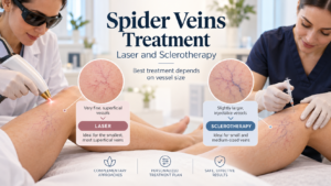
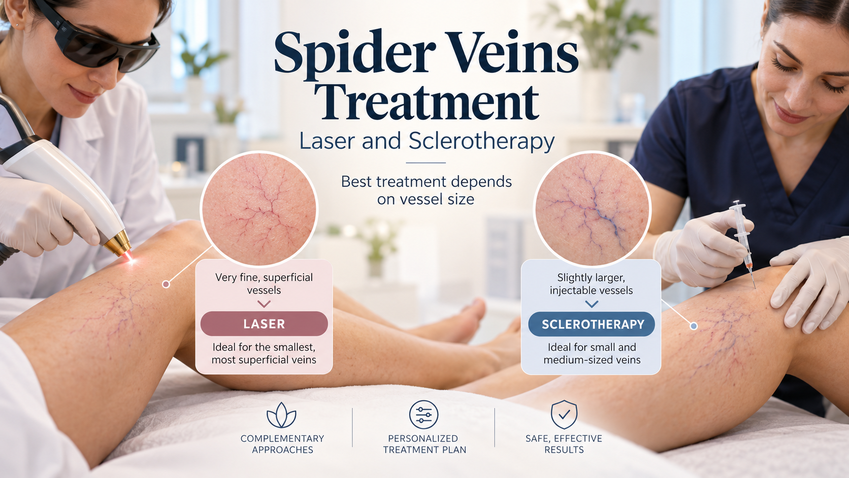
The most suitable treatment depends largely on the diameter of the blood vessel, its depth beneath the skin and whether it can be safely injected.
Understanding the Size of Spider Veins
Not all visible leg veins are the same size.
Spider veins, medically known as telangiectasias, generally measure less than 1 millimetre (mm) in diameter.
Slightly larger vessels, called reticular veins, generally measure between 1 and 3 mm. These veins are often blue or green and may supply blood to networks of smaller spider veins.
This difference matters because the two treatments work in different ways.
Sclerotherapy: An Effective Treatment for Larger, Injectable Veins
Sclerotherapy involves injecting a specialised medical solution directly into the affected vein through a very fine needle.
The solution, called a sclerosant, damages the inner lining of the vein. This causes the vessel to close. Over time, the body gradually breaks down the treated vessel, making it less visible.
Sclerotherapy is particularly useful for reticular veins and spider veins that are large enough to be accurately injected.
Its main advantages include:
Effectiveness in treating larger networks of superficial veins.
The ability to treat reticular or feeder veins that supply smaller spider veins.
Strong scientific evidence supporting its use.
The 2023 American venous disease guidelines recommend liquid or foam sclerotherapy for symptomatic spider and reticular veins. The 2022 European Society for Vascular Surgery guidelines also recommend sclerotherapy as the first-choice treatment for reticular veins.
However, sclerotherapy has a practical limitation: the treating clinician must be able to place the needle accurately inside the target vessel.
This becomes increasingly difficult as the vessel becomes smaller.
Vascular Laser: An Important Solution for Very Fine Spider Veins
Vascular laser therapy works without inserting a needle into the blood vessel.
Instead, the laser delivers concentrated light through the skin. The light is absorbed by haemoglobin, the pigment in red blood cells that carries oxygen.
This produces controlled heat inside the targeted vessel, damaging its walls and causing it to close. The body then gradually clears the treated vessel.
Different vascular lasers, including 532 nm KTP, 595 nm pulsed dye and 1064 nm Nd lasers, can be used depending on the vessel’s size, colour and depth, as well as the patient’s skin type.
Laser treatment is especially valuable for extremely fine spider veins that are difficult to inject.
Why the Diameter of a 30-Gauge Needle Matters
A 30-gauge needle, commonly written as 30G, has a nominal outside diameter of approximately 0.30 mm, with a standard manufacturing range of 0.298–0.320 mm according to ISO 9626:2016.
This means the needle itself is approximately three-tenths of a millimetre wide.
Now consider a spider vein measuring just 0.2 mm in diameter.
That vessel is narrower than the outside of a 30G needle. Accurately placing such a needle inside the tiny vessel is technically challenging and may damage the vessel or surrounding tissue.
For vessels smaller than approximately 0.3 mm, vascular laser therapy is therefore often the more practical and appropriate treatment option.
Laser treatment can close these fine vessels without requiring direct needle access.
Importantly, 0.3 mm is a practical guide rather than an absolute medical cutoff. Finer needles exist, and some small vessels can still be treated successfully with microsclerotherapy. Vessel depth, visibility, blood flow and clinician experience also influence treatment selection.
Nevertheless, the principle remains clear: when a vessel is too fine to inject reliably, vascular laser offers an important alternative.
What Does Scientific Research Tell Us About Effectiveness?
One of the most important scientific studies comparing these treatments is a 2021 Cochrane systematic review.
A systematic review examines results from several clinical studies to determine what the overall scientific evidence shows.
The Cochrane review examined 35 randomised clinical trials involving 3,632 participants.
It found that:
Sclerotherapy was effective at improving or clearing spider and reticular veins compared with placebo treatment.
Laser treatment and sclerotherapy showed no clear overall difference in effectiveness for spider vein clearance across the studies comparing them.
Laser treatment may cause less post-treatment skin pigmentation than sclerotherapy.
Combining laser with sclerotherapy may produce better vein clearance than sclerotherapy alone, although the evidence for this benefit was less certain and combined treatment could cause more discomfort.
These findings are important because they challenge the assumption that one technique must be superior to the other.
The European guidelines also describe clinical trials in which laser performed particularly well for smaller vessels, while sclerotherapy remained highly effective for larger reticular veins.
The correct conclusion is not that laser is always better, or that sclerotherapy is always better.
Both methods are effective, but their usefulness changes according to the characteristics of the blood vessel being treated.
How the Two Treatments Complement Each Other
Imagine a patient who has a network of fine red spider veins connected to a larger blue reticular vein.
Treating only the fine red vessels may leave the underlying feeder vein untreated.
A clinician may therefore choose to treat the larger reticular vein with sclerotherapy first.
The remaining fine spider veins can then be treated with vascular laser, especially when they are too narrow for accurate needle injection.
This approach uses each treatment where it is most suitable.
As a practical guide:
| Vessel diameter | Treatment commonly considered |
|---|---|
| Below approximately 0.3 mm | Vascular laser is often preferred because injection is technically difficult. |
| Approximately 0.3–1 mm | Laser or microsclerotherapy, depending on vessel characteristics and accessibility. |
| Approximately 1–3 mm | Sclerotherapy is generally preferred, particularly for reticular and feeder veins. |
These are useful clinical distinctions, not rigid treatment rules.
Scientific research also supports the potential benefits of combining the two approaches. The 2021 Cochrane review found that combining laser with polidocanol sclerotherapy may improve clearance compared with polidocanol alone. However, further high-quality research is needed to establish which treatment combinations work best.
Safety and Individual Assessment
Both treatments can produce side effects.
Sclerotherapy may cause temporary bruising, brown pigmentation, discomfort or the development of fine new blood vessels, known as telangiectatic matting. Rare but more serious complications can occur.
Vascular laser may cause temporary redness, swelling, discomfort, pigmentation changes or, occasionally, burns and scarring.
Some laser treatments, particularly Nd laser treatment, may be more painful than sclerotherapy.
The ideal treatment must therefore take account of more than vessel size alone. Skin type, vessel depth, symptoms, medical history and any underlying venous disease should also be considered.
Neither method guarantees permanent freedom from spider veins, because new vessels can develop over time.
Conclusion: The Right Treatment for the Right Vessel
The debate about whether laser or sclerotherapy is better for spider veins misses the most important clinical principle.
The aim is not to prove that one technology is superior, but to select the most appropriate treatment for the vessel being treated.
Sclerotherapy remains an established and highly effective approach for larger, injectable spider veins and reticular veins.
Vascular laser is particularly valuable for the finest vessels, including those narrower than the approximately 0.3 mm outside diameter of a 30G needle, where accurate injection becomes difficult.
When used appropriately, the two techniques can complement each other, allowing treatment of both larger feeder veins and smaller surface vessels.
The scientific evidence supports an individualised approach rather than a competition between techniques.
Ultimately, successful spider vein management depends not on choosing laser instead of sclerotherapy, or sclerotherapy instead of laser, but on understanding when to use each method—and when using both may offer additional benefit.
Scientific References
1. Cochrane Systematic Review (2021).
Nakano LCU et al. Treatment for telangiectasias and reticular veins. Cochrane Database of Systematic Reviews. Review of 35 randomised trials involving 3,632 participants.
Read the scientific review
2. Society for Vascular Surgery, American Venous Forum and American Vein and Lymphatic Society (2023).
Clinical Practice Guidelines for the Management of Varicose Veins of the Lower Extremities, Part II. Section 7.1 addresses laser and sclerotherapy for spider and reticular veins.
Read the clinical guidelines
3. European Society for Vascular Surgery (2022).
De Maeseneer MG et al. Clinical Practice Guidelines on the Management of Chronic Venous Disease of the Lower Limbs. European Journal of Vascular and Endovascular Surgery, 63, 184–267.
Read the published guidelines
4. International Organization for Standardization (2016).
ISO 9626:2016 — Stainless steel needle tubing for the manufacture of medical devices. Specifies a 30G needle’s outside diameter range of 0.298–0.320 mm.