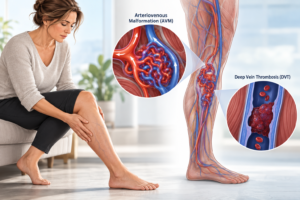
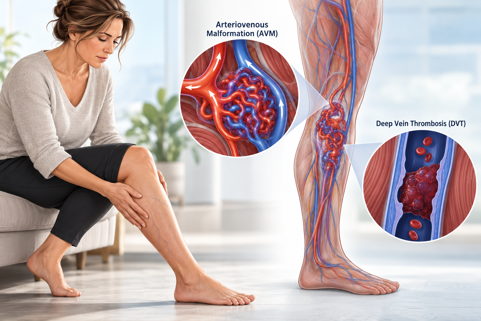

An arteriovenous malformation, or AVM, in the leg can sometimes exist alongside deep vein thrombosis (DVT), but the relationship is more complicated than simply saying that an AVM causes blood clots.
An AVM is a high-flow vascular malformation. Abnormal arteries connect directly with veins instead of sending blood through the normal network of tiny capillaries first. This causes unusually fast blood flow through the affected area and can enlarge both the feeding arteries and draining veins.
A DVT is different.
A deep vein thrombosis is a blood clot that forms inside one of the deep veins, most commonly in the calf, thigh or pelvis.
Someone with a leg AVM can develop a DVT, particularly when other problems affect normal venous drainage. However, DVT is not considered an inevitable or defining feature of an AVM.
In fact, current medical literature shows that the strongest vascular-malformation-related risk of venous thrombosis occurs in slow-flow venous and combined vascular malformations, rather than in isolated high-flow AVMs.
That distinction is important for anyone researching an AVM and blood clot risk.
What Is an Arteriovenous Malformation in the Leg?
To understand the problem, it helps to understand normal blood flow.
Normally:
Heart → artery → tiny capillaries → vein → heart
The arteries deliver oxygen-rich blood to the tissues.
Capillaries slow the blood down and allow oxygen and nutrients to move into muscles, skin, nerves and other tissues.
Veins then carry the blood back toward the heart.
In an AVM, some blood takes a shortcut:
Artery → AVM → vein
The normal capillary network is partly bypassed.
The abnormal group of connections is often called the nidus of the AVM.
Because there is very little resistance between the arteries and veins, blood can travel through the AVM very quickly. Peripheral AVMs are therefore known as fast-flow or high-flow vascular malformations.
Where Can an AVM Occur in the Leg?
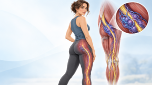
A lower-limb AVM can involve many different structures, including:
- the thigh
- buttock
- pelvis
- knee
- calf
- ankle
- foot
- muscles
- skin
- bone
- arteries
- veins
- tissue around nerves
Some AVMs are limited to a relatively small area.
Others can extend through several muscles or sections of the leg.
A complex AVM may have several feeding arteries and large draining veins.
The anatomy matters because a small superficial AVM can behave very differently from an extensive AVM involving the pelvis, thigh and deep venous system.
What Does a Leg AVM Feel Like?
The symptoms depend on the size, location and blood flow through the AVM.
Possible symptoms include:
- swelling
- pain
- aching
- heaviness
- warmth
- visible enlarged veins
- skin redness or discoloration
- a pulsing sensation
- a vibration or “buzzing” feeling
- bleeding
- skin ulcers
- difficulty walking
- muscle fatigue
- enlargement of the affected leg
- reduced function
Doctors may be able to feel a vibration over an AVM called a thrill.
A rushing sound heard with a stethoscope is called a bruit.
These findings happen because blood is moving rapidly through abnormal artery-to-vein connections.
Peripheral AVMs can cause venous hypertension, swelling and enlarged draining veins. More advanced lesions can lead to pain, ulcers, bleeding and tissue damage. Very extensive AVMs can place additional strain on the heart.
Why Can an AVM Make the Leg Swell?
Swelling is one of the most confusing symptoms because it can occur with both an AVM and a DVT.
In an AVM, high-pressure arterial blood enters the venous system much earlier than normal.
This can increase the pressure inside the draining veins.
Doctors call this venous hypertension.
Imagine connecting a high-pressure garden hose directly to a lower-pressure drainage pipe.
The drainage system suddenly has to handle much more blood, moving much faster than it normally would.
Over time, the draining veins can become:
- enlarged
- twisted
- overloaded
- more visible
- less efficient
The leg may then feel swollen, heavy, warm or uncomfortable.
This swelling can resemble the swelling caused by a DVT.
That means a person with a known AVM should not automatically assume that new swelling is “just the AVM.”
A new DVT may need to be ruled out.
What Is a DVT?
A deep vein thrombosis occurs when a blood clot develops inside a deep vein.
Most DVTs occur in the:
- calf
- thigh
- pelvis
A DVT can partly or completely block blood returning through the vein.
Typical symptoms include:
- new swelling in one leg
- calf or thigh pain
- tenderness
- warmth
- redness or skin discoloration
- more noticeable surface veins
Some DVTs cause few or no symptoms.
Can a Leg AVM Actually Cause a DVT?
The best answer is:
Possibly in some patients, but an isolated AVM should not automatically be considered a high-DVT-risk condition.
Modern research into blood clotting and vascular anomalies shows that slow-flow vascular malformations, especially venous malformations, are the group most clearly associated with abnormal clotting and venous thromboembolism.
AVMs are high-flow lesions and have different blood-flow characteristics.
However, several situations may bring AVMs and DVT together.
These include:
Abnormal or enlarged draining veins
An extensive AVM can dramatically alter the veins carrying blood away from the leg.
Some veins become enlarged because of the high volume of blood passing through them.
If other areas of the venous system are narrowed, obstructed or poorly developed, normal drainage may become more difficult.
Venous obstruction
A narrowed or blocked deep vein can cause blood to back up in the leg.
When venous obstruction and an AVM occur together, the combination can produce severe venous hypertension.
Previous DVT
A previous clot may permanently damage a deep vein.
Even after the clot has cleared, valves inside the vein may remain damaged.
This can cause post-thrombotic syndrome, with long-term swelling, heaviness, skin changes and sometimes ulcers.
Published reports have described patients with both iliofemoral AVMs and previous DVT who developed severe post-thrombotic problems.
Reduced mobility
Large or painful AVMs can make walking difficult.
Someone who moves less because of pain, surgery or disability may develop the same immobility-related DVT risk seen in anyone else.
Operations and interventional procedures
Vascular procedures can temporarily affect blood flow and may increase clotting risk in some patients.
The individual risk depends heavily on the type of malformation and procedure.
Other ordinary DVT risk factors
A patient with an AVM can still have the same risk factors for DVT as anyone else, including:
- previous DVT or pulmonary embolism
- major surgery
- prolonged bed rest
- long periods without moving
- serious injury
- pregnancy
- some estrogen-containing medicines
- cancer
- inherited blood-clotting disorders
- obesity
- smoking
- increasing age
The presence of an AVM therefore has to be considered as one part of the patient’s entire vascular anatomy and clotting risk profile.
AVM Versus Venous Malformation: The DVT Difference Matters
This is one of the most important parts of the discussion.
People often use the general term vascular malformation as if every malformation behaves in the same way.
They do not.
Venous malformation
A venous malformation involves abnormally formed veins.
Blood flow through these abnormal venous spaces is slow.
This slow movement of blood can encourage local clot formation and a condition called localized intravascular coagulopathy, or LIC.
Patients with large slow-flow venous malformations can therefore have a meaningful risk of superficial thrombosis, DVT and, in some circumstances, pulmonary embolism.
Arteriovenous malformation
An AVM has direct connections between arteries and veins.
Blood moves very quickly through the abnormal vessels.
AVMs are therefore considered fast-flow lesions.
The major problems associated with AVMs are more commonly related to:
- abnormal high blood flow
- venous hypertension
- pain
- swelling
- tissue damage
- ulceration
- bleeding
- enlargement of the lesion
- strain on the heart in very extensive disease
This does not mean a person with an AVM cannot develop DVT.
It means the clotting mechanism seen in slow-flow venous malformations should not automatically be applied to AVMs.
Can DVT Make an Existing AVM Worse?
Possibly.
The relationship can sometimes work in the other direction.
If a DVT blocks an important deep vein, pressure increases below the blockage.
The body may respond by developing alternative routes for blood flow.
There are published case reports and series in which AVM-like or arteriovenous communications became apparent after major venous thrombosis or obstruction.
Researchers have proposed that increased venous pressure, inflammation, low oxygen levels in tissues and the formation of new blood vessels may contribute to this process.
However, much of this evidence comes from unusual cases.
It would therefore be inaccurate to say that DVT routinely creates an AVM.
The important point is that DVT, venous obstruction and abnormal arteriovenous connections can sometimes interact in complicated ways.
Why Is DVT More Concerning in a Leg That Already Has an AVM?
An AVM already changes the normal circulation of the limb.
A DVT can then block part of the deep venous drainage system.
This can produce an even greater increase in venous pressure.
Possible consequences include:
- severe swelling
- worsening pain
- enlarged surface veins
- skin discoloration
- chronic inflammation
- skin thickening
- venous ulcers
- difficulty walking
- post-thrombotic syndrome
A 2025 report described seven patients who developed severe post-thrombotic syndrome after iliac DVT in limbs that also had iliofemoral arteriovenous malformations. These were unusual cases and should not be used to estimate the risk for every person with an AVM, but they demonstrate how serious the combination can become.
What Is Post-Thrombotic Syndrome?
After a DVT, the vein may never return completely to normal.
The clot can damage valves inside the vein.
These valves normally help blood travel upward toward the heart.
If they stop working properly, blood can pool in the leg.
This is called post-thrombotic syndrome, or PTS.
Symptoms may include:
- chronic swelling
- aching
- heaviness
- skin discoloration
- discomfort while walking
- enlarged veins
- skin ulcers
In someone who also has an AVM sending unusually high volumes of blood into the venous system, damaged or obstructed veins may have even more difficulty handling that blood flow.
Can an AVM and a DVT Cause the Same Symptoms?
Yes.
This is one reason diagnosis can be difficult.
Both conditions can cause:
AVM
- swelling
- pain
- visible veins
- warmth
- skin changes
DVT
- swelling
- pain
- visible surface veins
- warmth
- redness or discoloration
However, an AVM may also produce:
- pulsation
- a palpable thrill
- an audible bruit
- unusually warm skin
- long-standing enlargement of the limb
A DVT is more likely to cause a relatively sudden change, although this is not always the case.
Because symptoms overlap, a major new change in a limb with an AVM should be medically assessed rather than diagnosed by symptoms alone.
What Symptoms Should Raise Concern for DVT?
A person with a leg AVM should seek prompt medical assessment for a new or unexplained:
- increase in leg swelling
- swollen calf
- swollen thigh
- calf or thigh pain
- tenderness
- redness
- unusual warmth
- sudden heaviness
- change in skin colour
- sudden increase in visible veins
A DVT cannot reliably be diagnosed just by looking at the leg.
Some people with DVT have no obvious symptoms at all.
When Is It an Emergency?
The most dangerous complication of DVT is a pulmonary embolism, or PE.
This happens when part of a blood clot breaks away, travels through the bloodstream and becomes lodged in a blood vessel in the lungs.
Emergency symptoms can include:
- sudden shortness of breath
- chest pain
- chest pain that gets worse when breathing deeply
- coughing up blood
- rapid heartbeat
- feeling faint or dizzy
- collapse
A pulmonary embolism can be life-threatening and requires emergency treatment.
In Australia, call 000 for these symptoms.
How Do Doctors Check for DVT in Someone With an AVM?
The evaluation may be more complicated than in a person with normal vascular anatomy.
Doctors may use several tests.
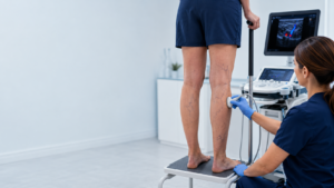
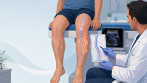
Duplex ultrasound
An ultrasound is one of the main tests used to investigate suspected DVT.
It can show whether a deep vein is compressible and whether normal blood flow is present.
With an AVM, Doppler ultrasound can also demonstrate:
- unusually fast arterial flow
- abnormal artery-to-vein shunting
- enlarged vessels
- arterialised flow within draining veins
This makes Doppler ultrasound useful for examining both the veins and the AVM’s blood-flow pattern.
D-dimer blood test
D-dimer is a substance produced as the body breaks down blood clots.
A D-dimer test can help doctors decide whether DVT is likely in certain clinical situations.
However, a high D-dimer does not prove that someone has a DVT.
Other illnesses and medical conditions can increase it.
Imaging is often required to identify whether a clot is actually present and where it is located.
MRI and MR angiography
MRI can show the full extent of a vascular malformation and its relationship to:
- muscles
- bones
- nerves
- arteries
- veins
MRI and magnetic resonance angiography can help distinguish high-flow AVMs from other types of vascular malformations.
CT angiography
CT angiography may provide detailed information about:
- feeding arteries
- draining veins
- the AVM nidus
- neighbouring anatomy
- venous obstruction
Catheter angiography
Digital subtraction angiography, or DSA, gives doctors a detailed moving picture of blood passing through the AVM.
It is particularly important when planning embolization.
Peripheral AVM reviews describe angiography as a critical method for understanding the architecture and flow pattern of the lesion.
What Should Imaging Look for in a Leg AVM With Possible DVT?
A thorough evaluation may need to answer more than simply:
“Is there an AVM?”
Important questions include:
- Where is the AVM nidus?
- Which arteries feed it?
- Which veins drain it?
- Are the draining veins unusually enlarged?
- Is the normal deep venous system open?
- Is there narrowing or obstruction of the iliac, femoral or popliteal veins?
- Is there an acute DVT?
- Has there been a previous DVT?
- Are there collateral veins?
- Is the AVM causing significant venous hypertension?
- Does the malformation involve muscle, bone, skin or nerves?
- Is there another type of vascular malformation present as well?
- Is the AVM isolated or part of a wider vascular syndrome?
These details can completely change the treatment strategy.
How Are Leg AVMs Treated?
Treatment depends on the AVM’s size, location, symptoms and vascular anatomy.
Not every AVM needs immediate treatment.
However, symptomatic or progressive peripheral AVMs can be challenging because they may recruit additional vessels and recur after incomplete treatment.
Embolization
Embolization is a major treatment for peripheral AVMs.
An interventional radiologist guides a small catheter through a blood vessel toward the abnormal connections.
Special materials are then used to block targeted parts of the AVM.
The goal is usually not simply to block one large feeding artery.
Effective treatment attempts to target the abnormal nidus or shunt itself.
If only a feeding artery is closed while the nidus remains active, the AVM may recruit other arteries and return.
Multiple procedures may sometimes be required.
Surgery
Surgery may be appropriate for selected AVMs, particularly when the lesion is:
- localized
- surgically accessible
- causing significant symptoms
- suitable for removal after embolization
Extensive AVMs growing through several tissues can be much more difficult to remove completely.
Combined treatment
Some patients receive embolization followed by surgery.
A multidisciplinary team usually decides which approach offers the best balance between controlling the AVM and preserving normal tissue and function.
How Is a DVT Treated When Someone Also Has an AVM?
A confirmed DVT is commonly treated with an anticoagulant, often called a blood thinner.
Anticoagulants reduce the ability of the clot to grow and decrease the chance of additional clots forming.
However, an AVM can also carry a risk of bleeding.
This creates an important clinical balancing act.
Doctors may need to consider:
- the location of the DVT
- whether there has been pulmonary embolism
- the size and location of the AVM
- whether the AVM has previously bled
- skin ulcers
- planned embolization or surgery
- other medications
- kidney function
- other clotting or bleeding disorders
For this reason, someone with an AVM should not start aspirin or anticoagulant medication simply because they are worried about DVT.
Blood-thinning treatment should be based on an individual medical assessment.
Does Everyone With a Leg AVM Need Blood Thinners?
No.
Having an AVM by itself does not mean a person should routinely take anticoagulants.
This is particularly important because AVMs are high-flow vascular abnormalities that can sometimes bleed.
The decision to use anticoagulation is different from the management of some large slow-flow venous malformations, where thrombosis and localized coagulopathy may be a major part of the condition.
Contemporary hematology reviews specifically emphasize the elevated venous-thromboembolism risk of slow-flow vascular malformations rather than applying the same risk profile to AVMs.
Anyone with both an AVM and a history of DVT may benefit from assessment by a vascular specialist and, in selected cases, a hematologist.
Can Treatment of a Vascular Malformation Cause DVT?
A blood clot is a recognized potential complication of some vascular-malformation interventions, although the risk varies considerably depending on the type of malformation and procedure.
Published studies of extremity vascular-malformation treatments have reported DVT among procedural complications, although it has not been a frequent complication in all groups.
Patients undergoing treatment should therefore discuss:
- their previous history of DVT
- previous pulmonary embolism
- family history of abnormal clotting
- medications
- mobility after treatment
- the anatomy of their deep veins
- whether thrombosis prevention is needed
with the treating team before the procedure.
Can Compression Stockings Help?
Compression garments can sometimes help control venous swelling in vascular conditions.
However, compression should be individualized in someone with a complex AVM.
The treating team should understand:
- arterial blood supply
- venous drainage
- the exact location of the AVM
- whether there is active DVT
- whether ulcers or tissue damage are present
before recommending a specific pressure or compression strategy.
This is especially important when abnormal arterial flow and venous obstruction exist together.
AVM, DVT and Virchow’s Triad
Doctors often explain DVT using something called Virchow’s triad.
It describes three broad reasons blood clots form:
1. Blood flow slows down
This may happen during prolonged immobility or when a vein becomes obstructed.
2. The blood vessel wall is injured
Surgery, trauma or previous thrombosis may damage the vessel.
3. The blood becomes more likely to clot
This can happen because of certain illnesses, medications or inherited clotting disorders.
A person with an AVM may have one or more of these additional factors even though the AVM itself is a high-flow lesion.
This explains why DVT risk should be assessed person by person, rather than assuming everyone with an AVM has the same risk.
Why a Multidisciplinary Vascular Anomalies Team Matters
Lower-extremity AVMs can involve several medical specialties at once.
Depending on the individual case, treatment may involve:
- interventional radiology
- vascular surgery
- vascular anomalies medicine
- diagnostic radiology
- hematology
- cardiology
- plastic or reconstructive surgery
- orthopedic surgery
- pain medicine
- physiotherapy
Specialist vascular-anomalies centres commonly use multidisciplinary care because the anatomy and treatment of these malformations can be complex.
When DVT or abnormal deep venous drainage is also present, the venous system needs to be considered alongside the AVM rather than treated as a completely separate problem.
Questions to Ask Your Specialist
Useful questions may include:
- Is my malformation definitely an AVM?
- Is it high-flow?
- Where is the AVM nidus?
- Which arteries feed it?
- Which veins drain it?
- Are my deep veins normal?
- Do I have venous obstruction or narrowing?
- Are any of my veins unusually enlarged?
- Do I have evidence of a current or previous DVT?
- Is my swelling caused by the AVM, venous hypertension, DVT or a combination?
- Do I have an increased personal risk of DVT?
- Do I need to see a hematologist?
- Would anticoagulation increase my bleeding risk?
- Is embolization appropriate?
- Would surgery ever be considered?
- Should the venous obstruction be treated?
- What symptoms should send me to the emergency department?
- What type of follow-up imaging will I need?
Frequently Asked Questions About Leg AVMs and DVT
Does an AVM cause blood clots?
Not usually in the same way as a venous malformation. AVMs are high-flow lesions. The strongest evidence for malformation-related venous clotting involves slow-flow venous and combined malformations. A patient with an AVM can still develop DVT because of abnormal venous anatomy, obstruction, previous DVT, reduced mobility, procedures or standard DVT risk factors.
Is a venous malformation the same as an AVM?
No.
A venous malformation contains abnormal veins and is usually slow-flow.
An AVM contains abnormal direct connections between arteries and veins and is high-flow.
The distinction can significantly affect symptoms, clotting risk and treatment.
Can an AVM cause one leg to swell?
Yes. An AVM can send large amounts of arterial blood directly into veins, producing venous hypertension and swelling.
How do I know whether swelling is from my AVM or a DVT?
You usually cannot know with certainty from symptoms alone. New unilateral swelling, pain, tenderness, warmth or skin discoloration should be assessed medically. Ultrasound is commonly used when DVT is suspected.
Can a DVT worsen an AVM?
DVT can obstruct normal venous drainage and greatly increase venous pressure in an affected leg. Case reports and small series have documented serious problems when DVT, venous obstruction and AVMs occur together.
Can an AVM cause varicose veins?
AVMs can cause enlargement of draining veins because unusually large volumes of blood are moving through them. These abnormal veins may look similar to varicose veins, although the underlying cause is different.
Can a DVT travel to the lungs?
Yes. A piece of a DVT can break away and travel to the lungs, causing a pulmonary embolism. This can be life-threatening.
Should a person with an AVM take aspirin to prevent DVT?
Not unless a doctor specifically recommends it. AVMs can bleed, and aspirin is not automatically appropriate simply because an AVM is present.
Does a high D-dimer mean I definitely have a DVT?
No. D-dimer can be elevated for several reasons. It can help doctors assess clotting risk, but it does not tell them where a clot is located and cannot diagnose DVT by itself.
What is the best scan for a leg AVM?
Doppler ultrasound, MRI/MR angiography, CT angiography and catheter angiography can all provide different information. MRI is particularly useful for defining the extent of a vascular malformation, while catheter angiography provides detailed information about its blood-flow architecture and is important for embolization planning.
Can a leg AVM be cured?
Some localized AVMs can potentially be controlled or removed very effectively. Extensive AVMs can be more difficult because incomplete treatment may allow the abnormal circulation to recur or recruit new vessels. Long-term specialist follow-up is often important.
The Most Important Takeaway
A leg AVM and a DVT are two different vascular problems, but they can sometimes occur together.
An AVM creates abnormal high-flow connections between arteries and veins.
A DVT is a blood clot inside a deep vein.
Current evidence does not support treating every isolated AVM as though it carries the same high clotting risk as a slow-flow venous malformation.
However, DVT risk may become more important when a person also has:
- abnormal or enlarged veins
- venous obstruction
- a previous DVT
- reduced mobility
- recent surgery or intervention
- another vascular malformation
- inherited or acquired clotting risks
Most importantly, new swelling or pain in a leg with an existing AVM should not automatically be blamed on the AVM.
A DVT may need to be excluded.
And when an AVM and DVT occur together, specialists need to consider both sides of the circulation: the abnormal arterial shunting and the ability of the deep veins to carry blood safely back toward the heart.
That is why complex lower-limb AVMs are best assessed by a multidisciplinary vascular anomalies team.
Medical Disclaimer
This article provides general educational information and is not a substitute for individual medical advice, diagnosis or treatment.
Seek prompt medical assessment for new one-sided leg swelling, pain, warmth or discoloration.
Seek emergency care for sudden shortness of breath, chest pain, fainting or coughing up blood because these can be symptoms of pulmonary embolism.
Key Medical Sources
This article was informed by the 2025 International Society for the Study of Vascular Anomalies classification, which defines AVMs as fast-flow vascular malformations with direct arterial-to-venous connections.
Current reviews of peripheral AVMs describe their characteristic venous hypertension, clinical progression, imaging and embolization-based management.
Recent hematology reviews distinguish the substantial venous-thromboembolism risk seen in slow-flow venous malformations from the physiology of high-flow AVMs.
Australian DVT and pulmonary-embolism symptom, diagnostic and emergency information was checked against Healthdirect Australia