
You eat a meal and suddenly feel exhausted, nauseated or light-headed. Standing makes your heart race. You have abdominal or pelvic pain, but scans and appointments never seem to produce a clear explanation.
For some people with Ehlers–Danlos syndrome or hypermobility spectrum disorder, this experience is painfully familiar.
These symptoms are often discussed separately. Abdominal pain goes to gastroenterology. Dizziness goes to cardiology. Pelvic pain goes to gynaecology or vascular medicine. But what if, in some patients, they are connected?
Researchers are exploring whether unusually flexible connective tissue may make certain blood vessels more vulnerable to compression—and whether that could contribute to symptoms associated with POTS, pelvic congestion and other conditions.
The idea is promising, but the research is still young. Here is what we know so far.
First, what is vascular compression?
Blood vessels do not float freely inside the body. They pass between organs, muscles, ligaments, bones and other vessels.
Sometimes, one of these surrounding structures presses on a blood vessel and narrows it. Blood may then struggle to move through the area, build up behind it or find another route through smaller veins.
That is the basic idea behind a vascular compression syndrome.
But there is an important catch: a compressed-looking vessel on a scan does not automatically mean that something is wrong. Some people have visible narrowing and no symptoms at all.
Doctors therefore need to look at the whole picture—not just the image.
Does the person’s pain match the location of the compression? Is blood flow actually affected? Are there enlarged veins or changes in pressure? Could something else explain the symptoms?
Those questions matter, especially before considering an invasive treatment.
Why might this affect people with EDS or hypermobility?
Connective tissue is often described as the body’s scaffolding. It helps support the skin, joints, blood vessels and internal organs.
In Ehlers–Danlos syndrome, or EDS, and hypermobility spectrum disorder, this tissue can be more flexible or fragile than usual.
That flexibility is usually discussed in relation to joints, but connective tissue is found throughout the body. Researchers suspect that reduced internal support may also allow organs and blood vessels to move more than expected.
This could change the angles between vessels or leave a vein with less protection from surrounding structures.
In other words, the same tissue laxity that contributes to joint instability may also affect the arrangement of structures inside the abdomen and pelvis.
It is a sensible theory. However, we still do not have large studies showing exactly how often this happens.
Which compression syndromes are researchers looking at?
Four conditions appear frequently in the literature.
Nutcracker syndrome
Despite the unusual name, Nutcracker syndrome has nothing to do with nuts.
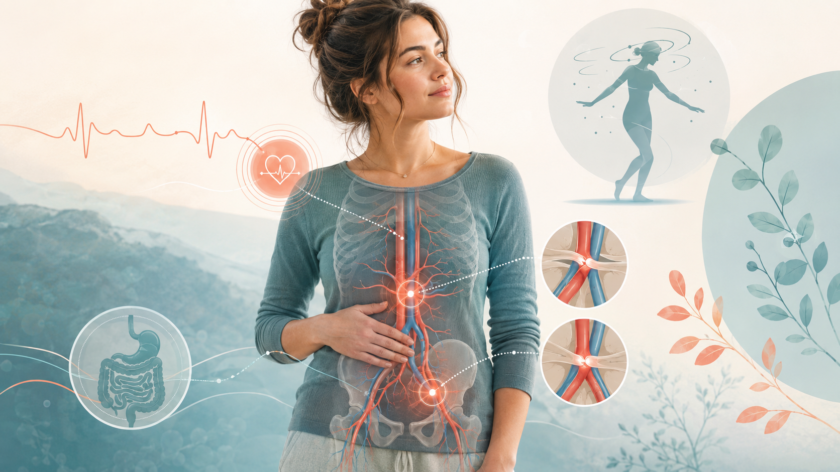
It usually happens when the vein draining the left kidney becomes squeezed between two major arteries.
That pressure can cause blood to back up and flow into veins around the pelvis, spine or reproductive organs.
Symptoms may include:
pain in the abdomen or side;
blood in the urine;
pelvic pain;
enlarged pelvic veins;
headaches;
symptoms that become worse while upright.
Some people also report difficulty eating or maintaining their weight.
May–Thurner syndrome
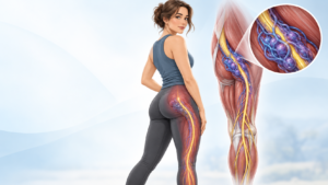
May–Thurner syndrome occurs lower in the pelvis. An artery presses against the vein that carries blood away from the left leg.
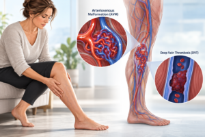
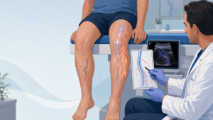
For some people, this causes no problems. For others, it may contribute to leg swelling, heaviness, varicose veins or blood clots.
It may also interfere with blood leaving the pelvis, which can contribute to chronic pelvic pain.
Median arcuate ligament syndrome
Median arcuate ligament syndrome, or MALS, involves pressure on an artery in the upper abdomen and sometimes on the nearby network of nerves.
People with symptomatic MALS often describe pain after eating. Nausea, vomiting and weight loss can follow because eating begins to feel like something to fear rather than enjoy.
One of the difficulties with MALS is that similar compression can sometimes be seen in people without symptoms.
Researchers are also unsure whether symptoms come mainly from reduced blood flow, irritated nerves or a combination of both.
Superior mesenteric artery syndrome
Superior mesenteric artery syndrome, or SMAS, is slightly different because it involves the intestine.
Part of the small intestine becomes trapped between two arteries, making it difficult for food to pass through normally.
This may cause pain after eating, early fullness, nausea, vomiting and weight loss.
Unfortunately, weight loss can make the problem worse. Fat normally provides some cushioning between the structures. As that cushion disappears, the space can become even narrower.
It can become a vicious cycle: eating causes pain, pain leads to eating less, and eating less increases the compression.
Where does blood flow come into the story?
This is where haemodynamics comes in.
The word sounds technical, but it simply refers to how blood moves through the body.
When you stand, gravity pulls blood down towards your legs, abdomen and pelvis. Your nervous system responds by tightening blood vessels and adjusting your heart rate. This helps push blood back towards the heart and brain.
The blood returning to the heart is called venous return.
If too much blood collects in the lower body, or if a compressed vein makes it harder for blood to return, the heart receives less blood with each beat.
The body may compensate by making the heart beat faster.
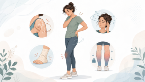
That can feel like:
dizziness;
a pounding heart;
weakness;
shakiness;
breathlessness;
difficulty thinking clearly.
This does not mean that every person with these symptoms has a vascular compression. It simply explains why a compression could potentially make orthostatic symptoms worse.
Is that how POTS fits in?
Possibly.
Postural orthostatic tachycardia syndrome, or POTS, is a disorder of the autonomic nervous system—the system that automatically manages heart rate, blood pressure, digestion and many other functions.
A key feature is an excessive increase in heart rate after standing.
POTS is commonly reported in people with EDS and hypermobility. One theory is that unusually stretchy veins allow more blood to pool in the lower body.
If a person also has a compressed vein in the abdomen or pelvis, that could create another obstacle to normal blood return.
Think of it less as one condition directly causing another and more as several factors piling on top of each other.
Flexible veins may encourage pooling. A compression may slow blood flow. The autonomic nervous system may then work overtime to compensate.
Some patients have reported an improvement in POTS symptoms after treatment for a vascular compression. That is encouraging, but it comes mainly from small studies and individual cases.
It does not mean that vascular surgery is a general cure for POTS.
Why can eating make everything feel worse?
Digestion needs blood.
After a meal, more blood is directed towards the stomach and intestines. Most people barely notice this shift.
But if your body already has difficulty keeping enough blood circulating to the heart and brain, that redistribution can leave you feeling light-headed, tired or unwell.
At the same time, conditions such as MALS and SMAS can directly cause pain or nausea after eating.
That means one meal can potentially trigger digestive symptoms and orthostatic symptoms together.
It is easy to see why patients—and sometimes clinicians—struggle to separate them.
And what about mast cell activation?
Mast cells are part of the immune system. They release chemicals such as histamine during allergic and inflammatory responses.
In mast cell activation syndrome, or MCAS, these chemicals are thought to be released too easily or at the wrong time.
People may experience flushing, itching, hives, headaches, reflux, abdominal discomfort, breathlessness, fatigue or faintness.
Histamine also widens blood vessels. In theory, that could encourage more blood pooling and make POTS symptoms worse.
Researchers have suggested that mast cell chemicals might also increase irritation around compressed vessels or nearby nerves.
However, this part of the story is still largely theoretical. There is not yet enough evidence to say exactly how MCAS and vascular compression influence one another.
What is pelvic congestion?
Pelvic congestion happens when blood collects in enlarged veins within the pelvis.
The pain is often described as heavy, dragging or aching. It may become worse after standing for a long time or later in the day.
Some people also experience pain during or after sex, bladder symptoms, vulval varicose veins or leg-vein problems.
Nutcracker syndrome and May–Thurner syndrome can both make it harder for blood to drain from the pelvis, so they may contribute to pelvic congestion in some patients.
Again, the phrase “in some patients” matters. Pelvic pain has many possible causes and deserves a broad assessment.
Why does diagnosis take so long?
Because almost every symptom in this area overlaps with something else.
Abdominal pain may come from a digestive disorder. Dizziness may come from POTS. Nausea may be blamed on medication, MCAS or gastroparesis. Pelvic pain may have gynaecological, muscular, neurological or vascular causes.
Once someone already has an EDS or POTS diagnosis, there is a risk that every new symptom will be placed under that label.
This is known as diagnostic overshadowing.
But the opposite can happen too. A narrowed vessel may appear on a scan and suddenly become the explanation for everything, even when the evidence is weak.
Good care needs to avoid both extremes.
The question should not simply be, “Is there a compression?”
It should be, “Is this compression affecting blood flow in a way that explains this person’s symptoms?”
How do doctors investigate it?
The starting point may be a Doppler ultrasound, CT scan or MRI.
A Doppler ultrasound looks at the speed and direction of blood flow. It is non-invasive, but the result can depend on the operator, the person’s position and the angle used during the scan.
CT and MRI can show how blood vessels relate to nearby organs and structures.
In more complex cases, specialists may use catheter-based imaging or intravascular ultrasound, which examines a vessel from the inside.
These tests can provide useful information, but they also carry risks. They should be chosen carefully rather than used simply because a patient has hypermobility and unexplained symptoms.
Are these tests safe for people with EDS?
For people with hypermobile EDS or HSD, the available reports suggest that invasive tests and procedures can often be performed safely.
The problem is that long-term evidence is limited.
Vascular EDS is a very different situation. In this subtype, arteries and internal organs can be extremely fragile.
Catheter procedures, high-pressure contrast injections and surgery may carry a much greater risk of vessel injury or rupture.
When vascular EDS is suspected, genetic assessment can be an important part of deciding which investigations are safe.
What does treatment involve?
There is no single treatment because each compression affects a different structure.
Some people may only need monitoring.
For others, improving nutrition and restoring weight can help rebuild the natural cushioning around the vessels and intestine.
More severe cases may involve:
placing a stent inside a narrowed vein;
moving a compressed vein;
releasing the ligament involved in MALS;
bypassing the obstructed section of intestine in SMAS;
treating enlarged or refluxing pelvic veins.
These treatments can be life-changing for carefully selected patients.
They can also fail to resolve symptoms, require further procedures or introduce new complications.
That is why finding a narrowing on a scan should be the beginning of a careful discussion—not an automatic ticket to surgery.
What does the research actually prove?
At the moment, less than many online discussions might suggest.
The literature review found a possible relationship between EDS or HSD, vascular compression, POTS, MCAS and pelvic congestion.
But much of the evidence comes from individual case reports, small patient groups and specialist clinics.
Only a small number of studies directly examined vascular compression in people with EDS or HSD.
We still do not know:
how common these syndromes truly are;
why one compression causes symptoms while another does not;
which patients are most likely to benefit from treatment;
whether treatment reliably improves POTS;
how stents and operations perform over many years;
how these conditions behave during pregnancy;
whether treating a compression changes MCAS symptoms.
So, there is a signal worth investigating—but not yet a complete answer.
What is the real takeaway?
People with EDS or hypermobility often spend years being told that their symptoms are unrelated, unexplained or simply part of their existing diagnosis.
The research suggests that, for some patients, vascular compression may be one missing piece of the puzzle.
It should be considered when symptoms follow a convincing pattern—for example, severe pain after eating, unexplained weight loss, blood in the urine, one-sided leg swelling, persistent pelvic pain or unusual orthostatic symptoms.
But it should not become the default explanation for every person with EDS or POTS.
The best approach is curious but cautious.
Listen to the patient. Look beyond a single diagnosis. Match the symptoms with the anatomy and blood-flow findings. Bring the right specialists together. And be honest about what the research does—and does not—yet tell us.
Sometimes the dots really are connected.
We just need better evidence before we can say exactly how.
This article is intended for general education and does not replace individual medical advice. Sudden leg swelling, chest pain, severe abdominal pain, ongoing vomiting or blood in the urine should be assessed by a healthcare professional.