
Phlebolymphoedema and venous insufficiency are two conditions that are closely linked. In this article, we will explore the connection between these two conditions and understand how they impact each other.
Phlebolymphoedema is characterised by the swelling of tissues caused by the accumulation of fluid and proteins in the affected area. Venous insufficiency, on the other hand, refers to the impaired functioning of the veins, particularly the ones responsible for returning blood to the heart.
The link between phlebolymphoedema and venous insufficiency lies in their shared underlying cause: a malfunctioning or damaged venous system. When the venous valves fail to function properly, it can lead to venous insufficiency, which in turn can result in phlebolymphoedema. Understanding this connection is crucial for effective diagnosis and treatment of both conditions.
As we delve deeper into this topic, we will explore the risk factors, symptoms, and treatment options associated with phlebolymphoedema and venous insufficiency. By shedding light on this connection, we hope to raise awareness and facilitate better management of these conditions for individuals facing them.
So, stay tuned for our in-depth exploration of the link between phlebolymphoedema and venous insufficiency.
Understanding phlebolymphoedema and chronic venous insufficiency
The Link Between Phlebolymphoedema & Venous Insufficiency:
Exploring the Connection
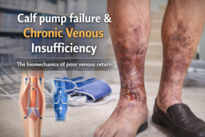
Chronic venous insufficiency (CVI) and phlebolymphedema are medical conditions that affect the circulatory system, particularly the veins and lymphatic vessels. CVI is a condition where veins in the legs fail to efficiently return blood from the extremities back to the heart due to damage or weakening of the vein walls and/or valves. Causes include venous valve dysfunction, deep vein thrombosis (DVT), and varicose veins. Symptoms include swelling, aching, leg heaviness, and skin changes. Complications include venous ulcers and superficial thrombophlebitis. Phlebolymphedema, a secondary lymphedema, occurs in conjunction with CVI and leads to impaired lymphatic drainage due to increased pressure in veins.
What Is the Connection Between Lymphedema and Phlebolymphedema?
Lymphedema and phlebolymphedema are related conditions characterized by fluid accumulation in tissue interstitial spaces, leading to swelling. Lymphedema is a chronic condition causing lymphatic fluid accumulation due to impaired drainage, often affecting the arms or legs. Phlebolymphedema is a specific type of secondary lymphedema arising from chronic venous insufficiency, which can cause increased pressure in veins and obstruct lymphatic vessels.
Both conditions are characterized by swelling, discomfort, skin changes, and potential complications like infections. Treatment approaches for both conditions often overlap, including compression therapy, manual lymphatic drainage, exercise, skincare, and lifestyle modifications. However, both conditions can lead to complications if left untreated. Effective management often requires a multidisciplinary approach involving healthcare professionals. Understanding the connection between lymphedema and phlebolymphedema is crucial for accurate diagnosis and effective management.
The Complex Interplay Between Venous and Lymphatic System
The relationship between phlebolymphoedema and venous insufficiency is complex and often cyclical. Venous insufficiency lays the groundwork for phlebolymphoedema to manifest, creating a cascade of symptoms that can be debilitating for patients. These symptoms may include:
Persistent Swelling: Swelling, medically known as edema, is a hallmark symptom of both phlebolymphoedema and venous insufficiency. This swelling is often accompanied by a feeling of heaviness or fullness in the affectedlimb.
Skin Changes: Over time, the compromised circulation in both conditions can lead to skin changes, such as hyperpigmentation, thickening, and even the development of venous ulcers.
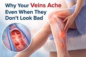
Pain and Discomfort: Chronic pain is a common complaint among individuals with phlebolymphoedema and venous insufficiency. This pain may be exacerbated by prolonged standing or sitting.
Reduced Quality of Life: The combined impact of these conditions can significantly diminish a patient’s quality of life, limiting mobility and causing emotional distress.
Symptoms and signs of phlebolymphoedema and venous insufficiency
Both phlebolymphoedema and venous insufficiency share common symptoms, such as swelling, aching, and heaviness in the affected limb. In phlebolymphoedema, the swelling is often accompanied by the leakage of lymphatic fluid, leading to a thickening and hardening of the skin. This can result in skin changes, such as discoloration, ulceration, and the development of venous stasis dermatitis.
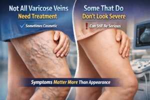
In venous insufficiency, the symptoms may vary depending on the severity of the condition. Early signs include swelling, particularly around the ankles, and the appearance of spider veins or varicose veins. As the condition progresses, individuals may experience pain, cramping, and aching in the affected limb. In severe cases, venous ulcers may develop, which can be difficult to heal and may require specialized wound care.
Causes of phlebolymphoedema and venous insufficiency
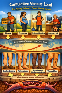
Phlebolymphoedema can be caused by a variety of factors, including genetic predisposition, trauma, surgery, infection, and obesity. In some cases, it may be a secondary condition that develops as a result of venous insufficiency. Venous insufficiency, on the other hand, is primarily caused by a malfunctioning or damaged venous system. This can be due to genetic factors, prolonged sitting or standing, pregnancy, obesity, or a history of blood clots. Understanding the underlying causes of these conditions is essential for effective diagnosis and treatment.
Pathophysiology of Lymphedema
Lymphedema is a chronic condition characterized by the accumulation of lymphatic fluid in interstitial spaces, leading to swelling and tissue changes. It is caused by impaired or damaged lymphatic systems, disrupting the normal flow of lymph fluid. The lymphatic system is essential for maintaining fluid balance, filtering waste products, and supporting the immune system.
It consists of lymphatic vessels, lymph nodes, lymphatic vessels, and pump mechanisms. The lymphatic system is equipped to handle a certain volume of lymphatic load and efficiently processes and returns lymph fluid to the bloodstream. However, lymphedema can occur due to surgical intervention, trauma or injury, or congenital abnormalities. Compromise of the lymphatic system impedes the normal drainage of lymph fluid, leading to swelling and tissue changes. Chronic inflammation, fibrosis, and thickening of skin and subcutaneous tissues can exacerbate the condition. Understanding the pathophysiology of lymphedema is crucial for developing effective treatment strategies.
Development of Phlebolymphedema (Secondary Lymphedema in Venous Insufficiency)
Phlebolymphedema, also known as secondary lymphedema, is a condition resulting from compromised blood and lymphatic circulation in the affected limb. It is a result of chronic venous insufficiency (CVI), where veins fail to efficiently return blood from the extremities to the heart. The condition develops due to damaged or weakened valves in veins, leading to increased pressure and fluid accumulation. Impaired lymphatic drainage further exacerbates the condition, causing fluid accumulation in interstitial spaces and compromising tissue health. The accumulation of fluid triggers an inflammatory response, leading to structural changes in the affected tissues and potentially leading to venous ulcers. The compromised circulation and immune function increase the risk of infections, particularly cellulitis, and the development of lipodermatosclerosis. Effective management and treatment are crucial for managing phlebolymphedema.
Pathophysiology of Chronic Venous Insufficiency
Chronic venous insufficiency (CVI) is a condition that arises from the impaired function of the venous system, particularly in the lower extremities. It is characterized by inadequate blood flow from the legs back to the heart. The pathophysiology of CVI involves a combination of factors, including venous valve dysfunction, venous hypertension, and altered microcirculation. Here’s a detailed explanation:
1. Venous Valve Dysfunction:
– CVI often originates from venous valve incompetence. Normally, one-way valves in the veins facilitate blood flow towards the heart and prevent backflow. In CVI, these valves become weakened or damaged, allowing blood to flow backwards (reflux) and pool in the lower extremities.
2. Venous Hypertension:
– The backflow of blood due to valve dysfunction leads to increased pressure in the veins of the lower limbs. This elevated pressure, known as venous hypertension, causes further dilation of the veins and impairs blood flow.
3. Venous Stasis and Pooling:
– As venous hypertension persists, blood accumulates in the veins, leading to venous stasis. This stagnant blood can cause the veins to dilate and become tortuous (varicosities), and can also lead to the development of spider veins.
4. Microcirculatory Changes:
– The increased pressure and altered blood flow dynamics in the veins can affect the microcirculation. This can lead to damage to small blood vessels and capillaries, resulting in tissue hypoxia (inadequate oxygen supply) and the accumulation of waste products.
5. Inflammatory Response:
– Chronic venous insufficiency triggers an inflammatory response in the affected tissues. This response includes the release of inflammatory mediators and recruitment of immune cells, which contribute to tissue damage and remodeling.
6. Tissue Fibrosis and Lipodermatosclerosis:
– Prolonged venous stasis and inflammation can lead to the deposition of fibrous tissue in the skin and subcutaneous tissues, a condition known as lipodermatosclerosis. This further compromises tissue integrity and can result in skin changes, including hyperpigmentation, induration, and ulceration.
7. Ulcer Formation:
– In severe cases of CVI, particularly when lipodermatosclerosis is present, chronic tissue damage and impaired wound healing can lead to the development of venous ulcers. These ulcers typically occur on the lower legs, often near the ankle, and can be slow to heal.
8. Complications:
– If left untreated, chronic venous insufficiency can lead to a range of complications, including venous ulcers, cellulitis (bacterial skin infection), and potentially deep vein thrombosis (DVT) due to altered blood flow dynamics.
Understanding the complex pathophysiology of chronic venous insufficiency is crucial for effective diagnosis and management. Treatment strategies typically aim to alleviate symptoms, improve venous function, and prevent complications, and may include lifestyle modifications, compression therapy, and, in some cases, surgical interventions.
Diagnosis and medical assessment
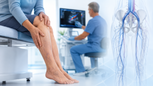
Diagnosing phlebolymphoedema and venous insufficiency involves a thorough medical assessment and examination. This may include a review of the individual’s medical history, a physical examination, and diagnostic tests such as ultrasound or venography. These tests can help determine the extent of the venous insufficiency and identify any underlying causes or complications. A proper diagnosis is crucial for developing an appropriate treatment plan and managing the symptoms effectively.
Why Does Early Detection of chronic venous disease Matter?
Early detection of chronic venous disease (CVD) is crucial for several reasons, including preventing progression, minimizing discomfort, preventing complications, improving quality of life, reducing healthcare costs, preserving skin health, maintaining work and daily activities, improving mental well-being, and enhancing overall health. Early detection allows for timely intervention, halting or slowing down the worsening of symptoms and complications.
It also helps alleviate discomfort and discomfort caused by CVD, such as pain, swelling, and cramping in the legs. Untreated CVD can lead to skin changes, ulcers, and blood clots, which can significantly impact a person’s overall health and quality of life. Addressing CVD early on can improve circulation, reduce blood clot risk, and contribute to better cardiovascular health. Regular check-ups with healthcare providers, especially for individuals with risk factors for CVD, can lead to timely diagnosis and treatment.
Non Surgical Treatment options for phlebolymphoedema and venous insufficiency
The treatment of phlebolymphoedema and venous insufficiency aims to alleviate symptoms, improve venous function, and prevent further complications. The specific treatment options may vary depending on the severity of the condition and the individual’s overall health. Conservative measures, such as compression therapy, elevation of the affected limb, and regular exercise, are often recommended as the first line of treatment. These measures help improve blood flow, reduce swelling, and alleviate discomfort.
In cases where conservative measures are ineffective, more invasive treatment options may be considered. This may include minimally invasive procedures, such as endovenous laser ablation or sclerotherapy, to seal off or remove the affected veins. In severe cases, surgical intervention may be necessary to repair or bypass damaged veins. The choice of treatment depends on several factors, including the extent of the condition, the individual’s overall health, and their treatment goals.
Effective management of phlebolymphoedema and venous insufficiency requires a multidisciplinary approach. This may involve:
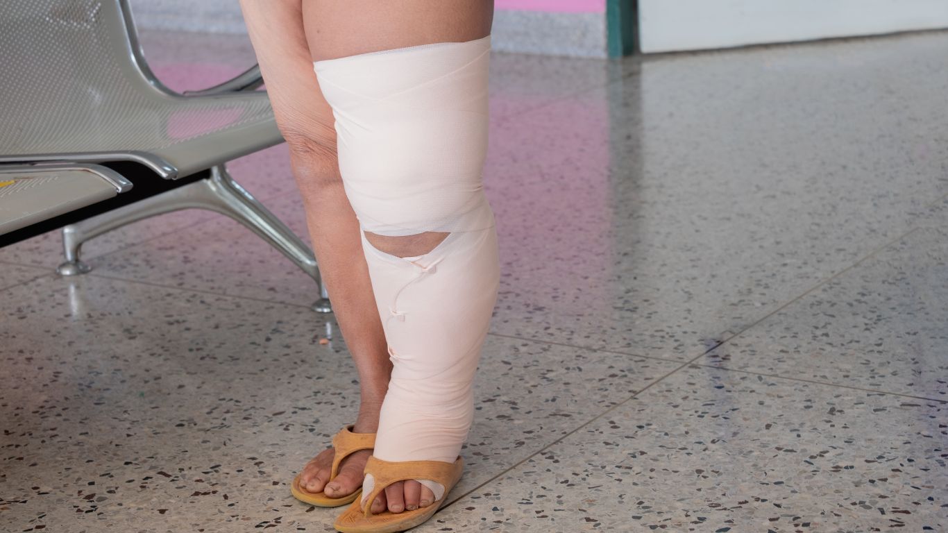
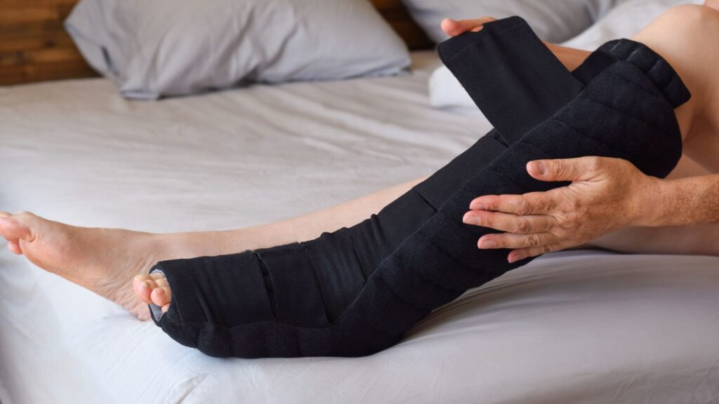
Compression Therapy: Compression garments can help improve blood and lymphatic flow, reducing swelling and discomfort.
Lifestyle Modifications: Lifestyle changes, such as regular exercise, maintaining a healthy weight, and avoiding prolonged periods of sitting or standing, are crucial in managing these conditions.
Medical Interventions: In severe cases, medical interventions such as minimally invasive procedures or surgery may be necessary to correct venous insufficiency.
Lymphatic Drainage Techniques: Manual lymphatic drainage and other specialized techniques can be employed to alleviate symptoms and improve lymphatic flow.
Minimally Invasive Treatment options for phlebolymphoedema and venous insufficiency
Minimally invasive treatment options for phlebolymphedema and venous insufficiency focus on improving venous circulation and managing lymphatic congestion. These interventions are effective in reducing symptoms and preventing complications associated with these conditions. Here are some common minimally invasive treatment options:
1. Endovenous Laser Ablation (EVLA):
– Description: EVLA uses laser energy to seal off abnormal veins. It is particularly effective for treating incompetent saphenous veins, which are often a source of venous insufficiency.
– Benefits: Minimally invasive, minimal discomfort, quick recovery, and high success rates in closing off problematic veins.
2. Radiofrequency Closure:
– Description: Similar to EVLA, radiofrequency closure uses thermal energy to close off problematic veins. It is particularly effective in treating large and tortuous veins.
– Benefits: Less discomfort compared to traditional vein stripping, shorter recovery time, and excellent success rates.
3. VenaSeal Closure System:
– Description: This system uses a medical adhesive to close off incompetent veins. It is an excellent option for patients who cannot tolerate heat-based treatments or have concerns about heat-related complications.
– Benefits: Minimally invasive, no need for tumescent anesthesia, and immediate return to normal activities.
4. Ambulatory Phlebectomy (Microphlebectomy):
– Description: Ambulatory phlebectomy involves the removal of varicose veins through tiny incisions. It is suitable for surface veins that are close to the skin’s surface.
– Benefits: Minimally invasive, does not require general anesthesia, and provides rapid relief from symptomatic varicose veins.
5. Sclerotherapy:
– Description: Sclerotherapy involves the injection of a sclerosing agent directly into the affected veins, causing them to collapse and eventually be reabsorbed by the body. It is particularly effective for treating smaller varicose veins and spider veins.
– Benefits: Minimally invasive, well-tolerated, and can be performed on an outpatient basis.
6. Lymphaticovenous Anastomosis (LVA):
– Description: LVA is a microsurgical procedure that creates direct connections between lymphatic vessels and nearby veins. It helps to redirect lymphatic fluid to the venous system, alleviating lymphedema symptoms.
– Benefits: Minimally invasive, reduces lymphatic congestion, and can lead to significant improvement in lymphedema.
7. Lymphatic Liposuction:
– Description: This specialized form of liposuction targets areas affected by lipedema, a condition often associated with lymphedema. It helps to remove excess fatty tissue and improve lymphatic flow.
– Benefits: Minimally invasive, can lead to significant improvement in symptoms, and enhances overall quality of life for patients with lipedema.
These minimally invasive treatments are typically performed on an outpatient basis, allowing for a faster recovery and reduced risk of complications compared to traditional surgical interventions. However, it’s important to note that the most appropriate treatment option will depend on the specific clinical presentation and needs of the patient.
Lifestyle changes and self-care tips for managing symptoms
In addition to medical interventions, individuals with phlebolymphoedema and venous insufficiency can benefit from making certain lifestyle changes and self-care tips. Maintaining a healthy weight, engaging in regular physical activity, and avoiding prolonged periods of sitting or standing can help improve blood flow and reduce symptoms. Practicing good skin hygiene, such as keeping the affected limb clean and moisturized, can also help prevent skin complications.
Wearing compression garments, as prescribed by a healthcare professional, can provide additional support to the affected limb and promote better circulation. It is important to follow the recommended guidelines for wearing and caring for compression garments to ensure their effectiveness. Additionally, individuals should be mindful of any changes in their symptoms and report them to their healthcare provider for timely intervention.
Research and advancements in phlebolymphoedema and venous insufficiency
Ongoing research and advancements in the field of phlebolymphoedema and venous insufficiency are paving the way for improved diagnosis and treatment options. Scientists and healthcare professionals are constantly exploring new technologies, medications, and interventions that can help individuals manage their symptoms more effectively. By staying updated with the latest research and advancements, healthcare providers can offer the best possible care to individuals with phlebolymphoedema and venous insufficiency.
Conclusion: Promoting awareness and understanding
Phlebolymphoedema and venous insufficiency are closely linked conditions that share a common underlying cause: a malfunctioning or damaged venous system. Understanding the connection between these conditions is essential for effective diagnosis and treatment. By recognizing the symptoms, addressing the underlying causes, and managing the symptoms effectively, individuals can experience significant improvements in their quality of life. It is important to promote awareness and understanding of these conditions to ensure timely intervention and support for those affected.