
Table of Contents
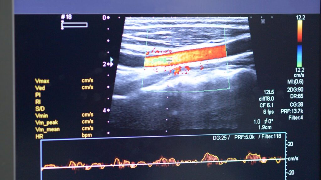
Duplex ultrasound has revolutionised the field of vascular imaging, providing a powerful tool for the diagnosis biomedical imaging and evaluation of various vascular conditions. This non-invasive imaging technique combines traditional ultrasound with both Doppler ultrasound and ultrasound waves. This combination provides detailed and real-time visualisation of blood flow within the arteries and veins. By utilising high-frequency sound, ultrasound waves, and advanced imaging technology, duplex ultrasound allows healthcare professionals to assess the structure, function, and blood flow characteristics of the blood vessels.
What is a Venous Duplex Ultrasound?
A venous duplex ultrasound scan is a non-invasive imaging test that combines a traditional ultrasound scan with Doppler ultrasound to assess the vessels in the body. This diagnostic medical sonography allows healthcare professionals to visualise the structure and function of the veins. Additionally, it allows to evaluate the direction and velocity of blood flow within them. By using sound waves and advanced imaging technology, ultrasound scan provides detailed information about the presence of blood clots, vein blockages, valve abnormalities, and other venous conditions. It is commonly used to diagnose and monitor conditions such as deep vein thrombosis (DVT), chronic venous insufficiency, varicose veins, and venous reflux. Ultrasound scan is a safe, generally painless,with a quick return to normal activities and an effective tool that plays a crucial role in the evaluation and management of venous disorders. It guides appropriate treatment decisions and optimising patient care.
Why would you need an ultrasound scan?
A venous duplex or ultrasound scan may be necessary for several reasons. It is commonly used to evaluate and diagnose various venous conditions, such as deep vein thrombosis (DVT), chronic venous insufficiency, varicose veins, and venous reflux. Here are some specific situations where a venous duplex or ultrasound scan may be needed:
1. Suspected Deep Vein Thrombosis (DVT):
If there is suspicion of a blood clot in the deep veins, a venous duplex or ultrasound scan can help confirm the diagnosis by visualising the affected veins and detecting the presence of a clot.
2. Varicose Veins or Venous Insufficiency:
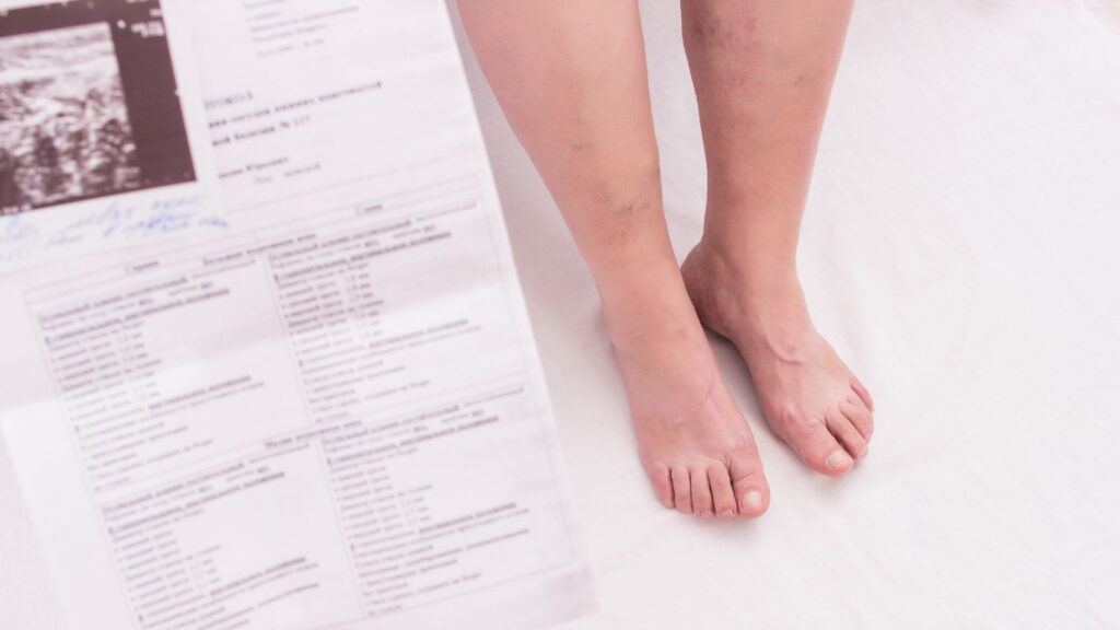
A venous duplex or ultrasound scan can assess the structure and function of the veins, helping to identify the underlying cause of varicose veins or chronic venous insufficiency, such as venous reflux or valve dysfunction.
3. Leg Swelling or Pain:
When a patient presents with unexplained abdominal ultrasound, leg swelling, pain, or other symptoms suggestive of venous disorders, an ultrasound scan can provide valuable information to determine the underlying cause of abdominal pain.
4. Preoperative Evaluation:
Prior to certain venous procedures or surgeries, a venous duplex or ultrasound exam may be performed to map out the veins, identify any obstructions or abnormalities, and guide the treatment planning.
5. Follow-up Monitoring:
For patients undergoing treatment for venous conditions, certain procedures such as sclerotherapy or endovenous ablation, regular venous duplex ultrasounds ultrasound examinations may be recommended to monitor treatment effectiveness and assess the progress of the condition.
What does a duplex ultrasound diagnose?
A duplex ultrasound is a non-invasive imaging technique that can diagnose and evaluate various conditions related to blood vessels pelvic organs and internal body structures. Specifically, it provides valuable diagnostic information about the structure and function of both arteries and veins. Here are some of the conditions medical issues that a duplex or ultrasound exam can diagnose:
1. Arterial Diseases:
Duplex, diagnostic ultrasound device that can identify arterial blockages or stenosis, which can indicate peripheral artery disease (PAD), carotid artery disease, or other arterial disorders. It is diagnostic ultrasound helps evaluate blood flow, detect any abnormalities in the arterial walls, and assess the severity of the disease.
2. Venous Diseases:
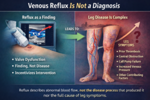
Duplex ultrasound is commonly ultrasound used to diagnose venous disorders such as deep vein thrombosis (DVT), chronic venous insufficiency, varicose veins, and venous reflux. It is ultrasound can visualize the veins, detect blood clots, assess venous flow and valve, imaging test for function, and identify any structural abnormalities or obstructions.
3. Vascular Anomalies:
Duplex ultrasound can identify vascular malformations, such as arteriovenous fistulas or vascular tumors. It helps determine the size, location, and characteristics of these anomalies, guiding further management and treatment decisions.
4. Transplants and Dialysis Access:
Duplex ultrasound is used to see blood flow help evaluate the patency and functionality of vascular grafts and fistulas used for dialysis access. It assesses blood flow, detects any narrowing or blockages, and helps ensure proper functioning of these vascular access sites.
5. Vascular Trauma:
Duplex ultrasound plays a crucial role in diagnosing and monitoring vascular injuries, such as arterial or venous dissections, pseudoaneurysms, or arteriovenous fistulas resulting from trauma. It helps assess the extent of the injury, identify any associated complications, and guide treatment strategies.
Overall, duplex ultrasound is a versatile diagnostic tool that provides valuable insights into the structure and function of blood vessels. It enables early detection, accurate diagnosis, and effective management of various vascular conditions, ultimately improving patient care and outcomes.
How Does Ultrasound Scans Work?
Ultrasound scans, most ultrasound scans or exams also known as sonography. It utilises high-frequency sound waves to create images of the internal structures of the body. Here’s a simplified explanation of how the most ultrasound exams or scans also called sonography work:
1. Sound Wave Generation:
A handheld ultrasound device, called ultrasound machine, a transducer emits high-frequency sound waves into the body. The transducer contains crystals that vibrate when an electric current passes through them, producing sound waves.
2. Sound Wave Propagation:
The sound waves travel through the body, penetrating the tissues. They encounter different types of soft tissues, such as organs, blood vessels, or bones, which can reflect or absorb the sound waves to create varying degrees.
3. Reflection and Echo Formation:
When the sound waves encounter a boundary between tissues of different densities, such as the boundary between fluid and solid tissues in human ear, some of the sound waves bounce back (reflect) while others continue to travel deeper into the inside of the human hearing body. These reflected sounds are called echoes.
4. Echo Reception:
The transducer also acts as a receiver, detecting the echoes produced by the reflected sound waves. The echoes are converted into electrical signals and sent to a computer.
5. Image Formation:
The computer processes the received signals and uses the timing and strength of the echoes to produce images to create a detailed image. The echoes from different tissues internal body structures and organs are displayed as varying shades of gray on computer screen or the ultrasound monitor. Dense or solid structures appear brighter, while fluid-filled or less dense structures appear darker.
6. Real-Time Imaging:
Ultrasound scans are typically performed in real-time, meaning that the images are generated instantly and continuously updated on the monitor as the transducer moves across the inside of the body. This allows the sonographer or healthcare professional to observe the structures in motion, such as the beating of the heart or the flow of blood in vessels.
Ultrasound-Guided Procedures
Ultrasound guidance for procedures refers to the use of ultrasound imaging during medical interventions ultrasound examinations imaging tests or treatments to provide real-time visualization and guidance. Here’s an overview of how this type of ultrasound imaging and guidance works:
1. Preparing for the Procedure:
To prepare for an ultrasound procedure, the patient is positioned appropriately on exam table, and the area to be treated or accessed during outpatient procedure is cleaned and sterilized.
2. Ultrasound Equipment Setup:
Ultrasound procedures coupled with ultrasound exams require ultrasound probe to remain sterile by covering with a sterile sheath or gel to ensure aseptic conditions. The ultrasound probe is then connected to an ultrasound machine.
3. Image Acquisition:
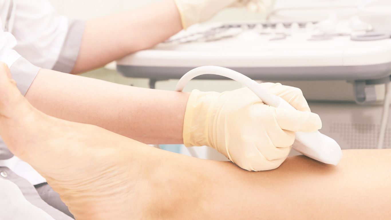
The healthcare professional applies ultrasound gel to the area of interest and places the probe on the skin. By moving the ultrasound probe over the targeted area, real-time ultrasound images are obtained.
4. Needle or Instrument Placement:
Using the ultrasound image as a guide, the healthcare professional can visualize the target area and guide the placement of a needle, catheter, or other instruments with precision and accuracy. The needle or instrument is inserted into the body through a small incision or puncture site.
5. Real-Time Monitoring:
Throughout the procedure, ultrasound technician or the healthcare professional continuously monitors the ultrasound image to ensure the correct placement and trajectory of the needle or instrument. Adjustments can be made in real time to optimize accuracy and avoid important structures.
6. Confirmation of Placement:
Once the needle or instrument is in the desired location, additional imaging tests further testing may be performed to confirm its proper placement and ensure that the intended target has been reached.
Ultrasound guidance offers several advantages, including increased accuracy, reduced risk of complications, and improved patient safety. It allows for precise targeting of specific areas or structures, even in challenging anatomical locations. Common procedures that benefit from this type of ultrasound guidance include biopsies, injections, aspirations, catheter placements, and drainage procedures.
By providing real-time visualization diagnostic ultrasound, and guidance, ultrasound guidance enhances the accuracy and success of various medical procedures, improving patient outcomes and minimizing risks.