
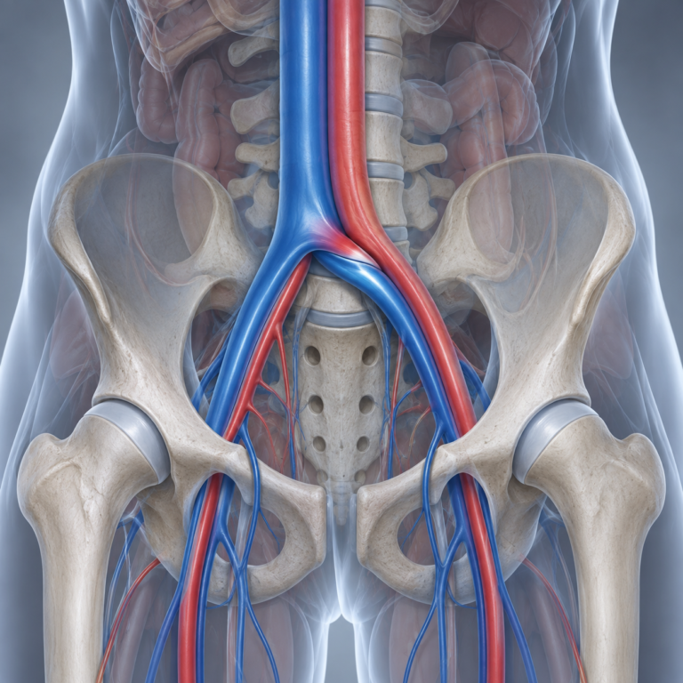
May-Thurner syndrome occurs when the right common iliac artery compresses the left common iliac vein, reducing blood flow return of the left leg. Dedicated clinical and ultrasound assessment can help confirm the diagnosis and guide appropriate treatment.
Detailed history, examination and vascular ultrasound
tailored to your specific findingsand goals.
experienced team in complex vacular management
conservative and interventional options when appropriate
May-thurner syndrome, also known as iliac vein. compression syndrome, occurs when the right common iliac artery compresses the left common iliac vein against the spine.
This can reduce blood flow from the left leg, leading to symptoms such as leg swelling, pain, heaviness and an increased risk of DVT. May-Thurner syndrome is recognised cause of unexplained left sided leg symptoms
The right common iliac artery compresses the left common iliac vein, reducing blood flow from the left leg back to the heart.
The narrowed vein can predispose to blood clot formation, particularly iliofemoral DVT (clot in the pelvic and upper thigh)
May lead to ongoing leg swelling pain, heaviness, visible veins or skin changes if not diagnosed and managed appropriately.
Symptoms can vary depending on whether the nerve, vein or artery is affected. Common symptoms include:
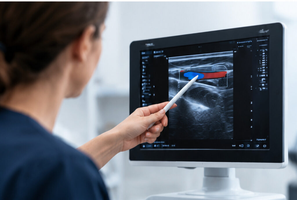
We take a comprehensive approach, combining your history and symptoms with a detailed clinical examination and targeted vascular ultrasound. In selected cases, further imaging such as CT or MR venography may be recommended to confirm the diagnosis and assess the degree of compression.
Anatomical compression: The right common iliac artery crosses over and compresses the left common iliac vein against the lumbar spine.
Repeated arterial pulsation: The artery’s continuous pulsations can irritate and damage the inner lining of the vein.
Vein narrowing: Over time, scar-like tissue or fibrous spurs may develop, further narrowing the vein.
Reduced blood flow: The narrowed vein restricts blood returning from the left leg, potentially causing venous congestion.
Increased risk of blood clots: Sluggish blood flow can increase the risk of deep vein thrombosis (DVT).
Treatment depends on the type and severity of your symptoms, We develop a personalised plan based on your diagnosis, lifestyle and goals.
Activity modification
physiotherapy or posture correction
Compression stocking
Anticoagulation
A catheter is inserted into the left iliac vein. When it’s in the correct position, a small mesh tube (stent) is put in place in the vein to keep it open
For select cases, we work closely with surgeons to organise surgical procedure reducing compression on the left iliac vein by moving the right iliac artery.
A member of our team will be in touch soon. Please, allow 24 hours to receive a response to your online form submission.

Something isn’t Clear?
I will be more than happy to answer all of your questions.