

“My scan was normal, so why do my legs still feel wrong?”
It is frustrating to be told that your duplex ultrasound does not explain your symptoms when your legs clearly feel heavy, swollen, tight, painful or visibly abnormal.
Many patients expect a venous scan to give a simple yes-or-no answer. Sometimes it does. If the scan shows clear great saphenous vein reflux, small saphenous vein reflux, a perforator problem or a deep vein clot, the next step is usually easier to understand.
But venous disease is not always that tidy.
Some people have symptoms that feel very much like varicose veins or venous congestion, yet their first duplex scan shows only mild reflux, no significant reflux, or findings that seem too small to explain the amount of discomfort. That does not mean the symptoms are imaginary. It means the diagnosis may need a wider lens.
A duplex ultrasound is an important test, but it is one piece of the clinical picture.
What duplex ultrasound is very good at showing
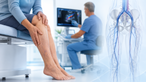
A leg venous duplex ultrasound is usually the first-line investigation for suspected varicose veins and chronic venous insufficiency. It can assess how blood is flowing through the veins, whether the valves are allowing backward flow, and whether there is evidence of acute or previous deep vein thrombosis.
It is particularly useful for identifying:
- Superficial venous reflux
- Great saphenous vein reflux
- Small saphenous vein reflux
- Accessory vein reflux
- Some perforator vein problems
- Deep vein thrombosis
- Some signs that may suggest deeper venous obstruction
For many patients, this test gives the information needed to plan treatment.
However, the quality of the result depends on the right question being asked, the right protocol being used, and the patient being scanned in the right position. A reflux study performed lying down may not show the same findings as a properly performed standing or weight-bearing reflux study. Venous symptoms are often worse when gravity is involved, so technique matters.
Why symptoms and duplex findings may not match
When duplex findings do not explain symptoms, there are several possibilities. Some are technical. Some are anatomical. Some are related to other conditions that can mimic venous disease.
1. The reflux may be position-dependent
Venous reflux is often more obvious when a person is standing. If the scan is performed mainly while lying flat, reflux may be underestimated.
This is one reason a patient can be told that the scan is “normal” while still having classic symptoms such as heaviness, aching, throbbing, swelling or visible veins that worsen during the day.
A good venous reflux assessment should match the patient’s symptoms, visible vein pattern and clinical history. The scan should not be interpreted in isolation.
2. The problem may be higher up in the pelvis or abdomen
A standard leg duplex scan focuses on the veins in the leg. It may not fully assess the pelvic veins, iliac veins or inferior vena cava.
This matters because some patients have venous outflow obstruction or venous compression higher in the body. Blood may struggle to drain from the leg or pelvis, even when the superficial leg veins do not show severe reflux.
Examples include:
- Iliac vein compression
- May-Thurner syndrome
- Non-thrombotic iliac vein lesions
- Post-thrombotic iliac vein obstruction
- Pelvic venous congestion
- Ovarian or internal iliac vein reflux
- Nutcracker syndrome in selected pelvic venous presentations
These conditions can create symptoms that overlap with varicose vein disease.
Venous compression syndrome: when the blockage is not in the visible veins
Venous compression syndrome occurs when a vein is narrowed or compressed by nearby structures. One of the best-known examples is May-Thurner syndrome, where the left common iliac vein is compressed in the pelvis.
This can affect venous drainage from the leg. Some patients develop left leg swelling, heaviness, aching, tightness, recurrent varicose veins or a history of deep vein thrombosis. Others have more subtle symptoms and no obvious clot.
The tricky part is that a routine leg duplex ultrasound may not fully show the compressed pelvic vein. It may show indirect clues, such as abnormal flow patterns, but the actual compression can be difficult to see.
That is why some patients need additional imaging when symptoms strongly suggest outflow obstruction.
Symptoms that may suggest a deeper venous issue
Not every aching leg is a venous compression problem. But certain patterns should raise the question, especially when the duplex result does not fit the symptoms.
A deeper venous or pelvic venous cause may be considered when there is:
- One-sided leg swelling, especially left-sided
- Leg heaviness or tightness that worsens through the day
- Symptoms worse with standing and improved with elevation
- Varicose veins in the groin, upper thigh, buttock, vulval, scrotal or lower abdominal area
- Recurrent varicose veins after previous treatment
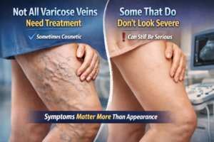
- Discomfort that seems out of proportion to the visible veins
- A history of deep vein thrombosis
- Pelvic heaviness or pelvic pain, especially worse with standing
- Symptoms that flare around menstruation or after pregnancy
- Leg swelling without significant superficial reflux
- Skin changes or venous ulcers with limited superficial findings
The key point is not that these symptoms confirm compression. They do not. The point is that they may justify a more complete venous assessment.
Could the symptoms be from something other than veins?
Yes. This is just as important.
When duplex findings do not explain symptoms, it is possible that the symptoms are not primarily venous. Several conditions can feel similar to varicose vein or venous congestion symptoms.
These include:
Lymphedema
Lymphedema can cause chronic swelling, heaviness, tightness and skin thickening. It often affects the foot and toes more than typical venous swelling.
Lipedema
Lipedema usually causes painful, heavy, symmetrical enlargement of the legs and can be mistaken for venous disease. The feet are often relatively spared.
Arterial disease
Arterial disease can cause exertional calf, thigh or buttock pain that improves with rest. This is different from venous heaviness, but patients may describe both as “leg pain.”
Nerve-related pain
Sciatica, spinal stenosis and peripheral neuropathy can cause aching, burning, tingling, numbness or heaviness. These symptoms may coexist with visible veins but not be caused by them.
Musculoskeletal problems
Hip, knee, back, tendon and muscle problems can create leg discomfort that overlaps with venous symptoms.
This is why the best assessment starts with the person, not the scan. The pattern, timing, triggers and location of symptoms matter.
What to do when your duplex scan does not match your symptoms
If your symptoms are persistent, worsening or affecting your quality of life, it is reasonable to ask for a more complete review.
A vascular specialist may consider:
1. Reviewing the original duplex report
Not all venous ultrasound reports provide the same level of detail. A specialist may look at whether the scan assessed:
- The deep veins
- The superficial veins
- The great and small saphenous veins
- Accessory veins
- Perforator veins
- Reflux times
- Vein diameters
- Flow phasicity
- Signs of previous thrombosis
- The iliac veins or IVC, if clinically relevant
Sometimes the issue is not that the scan is wrong. It may simply be incomplete for the symptoms being investigated.
2. Repeating the duplex with a venous reflux protocol
A repeat scan may be helpful if the original study was not performed standing, did not include a complete reflux map, or did not assess the area where symptoms and visible veins are most prominent.
3. Looking for signs of pelvic or iliac vein involvement
If symptoms suggest venous outflow obstruction, further assessment may include iliac vein and inferior vena cava ultrasound where technically possible.
4. Considering CT venography or MR venography
CT venography and MR venography can show anatomy that a leg duplex may not fully capture. These tests may help identify compression, narrowing, pelvic venous disease or other causes of swelling.
5. Considering venography and intravascular ultrasound in selected cases
Intravascular ultrasound, often called IVUS, is an invasive test performed from inside the vein. It may be used when a specialist needs detailed information about the severity and exact location of iliac vein narrowing, particularly if treatment such as venous stenting is being considered.
This is not a routine test for every patient with leg pain. It is usually reserved for carefully selected cases.
Why “mild reflux” may still matter in some patients
Sometimes a report says “mild reflux” and the patient is told it is not important. That may be true. But mild findings can still matter depending on the patient’s symptoms, vein pattern and previous history.
For example, a small amount of reflux in the wrong location may contribute to visible varicose veins. Or mild superficial reflux may coexist with deeper outflow obstruction, where the superficial veins are acting as overflow pathways.
This is why a good venous consultation should connect three things:
- What the patient feels
- What the clinician sees
- What the imaging shows
If those three do not line up, the next step is not guesswork. It is a more targeted investigation.
When to seek urgent medical care
Most varicose vein and chronic venous symptoms are not emergencies. However, urgent assessment is needed if you develop:
- Sudden one-sided leg swelling
- New severe calf or thigh pain
- Redness, heat and tenderness along a vein
- Shortness of breath or chest pain
- Coughing blood
- Sudden bleeding from a varicose vein
- A rapidly worsening wound or signs of infection
These symptoms may indicate a clot, bleeding complication, infection or another urgent condition.
Questions to ask your vascular specialist
If your scan does not explain your symptoms, these questions can help move the conversation forward:
- Was my scan a full venous reflux study?
- Was I scanned standing or lying down?
- Were the pelvic veins, iliac veins or IVC assessed?
- Do my symptoms suggest reflux, obstruction, compression or another cause?
- Could my visible veins be coming from pelvic or abdominal venous pressure?
- Should we consider CT venography, MR venography or another imaging test?
- Could lymphedema, lipedema, nerve pain or arterial disease be contributing?
- What findings would change the treatment plan?
A normal scan should not end the conversation if symptoms are ongoing and clinically significant.
The bottom line
Duplex ultrasound is one of the most useful tests in venous medicine, but it does not answer every question.
If your duplex findings do not explain your varicose vein symptoms, the next step is not to dismiss the symptoms. It is to ask whether the scan was complete, whether the problem may be higher up in the pelvis or abdomen, and whether another condition may be mimicking venous disease.
Persistent leg swelling, heaviness, pelvic pressure, recurrent varicose veins or symptoms that worsen with standing deserve a careful, whole-person assessment.
In many cases, the answer is not “nothing is wrong.” The answer may be, “we need to look in the right place.”
FAQs
Can I have varicose vein symptoms with a normal duplex ultrasound?
Yes. A duplex ultrasound may be normal or show only mild findings even when symptoms are real. The cause may be position-dependent reflux, pelvic venous disease, iliac vein compression, lymphedema, nerve pain, arterial disease or a musculoskeletal condition.
Can duplex ultrasound miss May-Thurner syndrome?
A routine leg duplex may miss iliac vein compression because the iliac veins sit deep in the pelvis and can be technically difficult to assess. If symptoms suggest compression, additional imaging may be needed.
What symptoms suggest venous compression syndrome?
Possible symptoms include one-sided leg swelling, heaviness, tightness, aching, pelvic pressure, recurrent varicose veins, groin or lower abdominal varices, and symptoms that worsen with standing and improve with elevation.
What test shows venous compression syndrome?
Depending on the situation, assessment may include iliac/IVC duplex ultrasound, CT venography, MR venography, catheter venography or intravascular ultrasound. The right test depends on symptoms, risk factors and whether treatment is being considered.
Should I ignore varicose vein symptoms if my scan is normal?
No. A normal scan is reassuring, but it does not always explain persistent symptoms. If symptoms continue, worsen or affect daily life, a vascular review may be appropriate.
Who should review unexplained venous symptoms?
A vascular surgeon, phlebologist, interventional radiologist or venous specialist can review symptoms, examine the pattern of veins and decide whether further imaging is needed.