

Hemodynamics of Varicose Veins:
Why Veins Fail and What Really Happens Inside
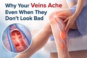
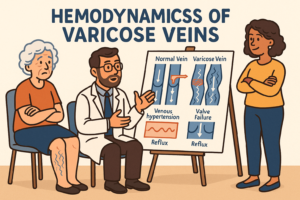
Varicose veins are often thought of as a cosmetic concern—bulging, twisted blue lines on the legs. But beneath the surface lies a complex story of venous pressure, valvular failure, and altered blood flow. These hemodynamic changes are what truly define the condition and drive both symptoms and progression.
This blog explores the why behind varicose veins: what happens to blood flow, how venous valves fail, and why some veins dilate while others don’t.
The Basics: How Healthy Venous Hemodynamics Work
After oxygen is delivered to the tissues, blood must return to the heart to be re-oxygenated and recirculated. This return trip happens through the veins, which carry deoxygenated, nutrient-depleted blood back toward the central circulation. In the legs, this task is especially challenging because blood must travel upward, against gravity, often while the body is standing or sitting still. To overcome this, the venous system relies on specialized structures and coordinated mechanisms that keep blood moving efficiently back to the heart.
a. The Calf Muscle Pump – The “Peripheral Heart”
The calf muscles act as a powerful pump. When they contract during walking or movement, they squeeze the deep veins, propelling blood upward. When the muscles relax, the veins refill from below. This pumping action is essential for lowering venous pressure in the legs and preventing blood from pooling.
b. Venous Valves – Why They Exist and How They Work
Because gravity constantly pulls blood downward, veins are equipped with one-way valves—thin, flap-like structures inside the vein that open to allow blood to move upward and snap shut to prevent it from flowing backward (reflux).
Valves are found throughout:
Superficial veins
Deep veins
Perforator veins (which connect the two systems)
Their main functions are to:
Maintain unidirectional flow toward the heart
Keep venous pressure low
Prevent blood pooling in the lower legs
Distribute blood properly between venous compartments
Without valves, blood would stagnate, venous pressure would rise, and veins would progressively dilate and fail.
c. Superficial, Deep, and Perforator Veins – A Coordinated Hemodynamic Network
The leg’s venous system is divided into three interconnected components, each with a distinct role:
Superficial veins (e.g., the great and small saphenous veins) lie just beneath the skin and drain blood from the skin and subcutaneous tissues.
Deep veins run between the muscles and handle the majority of venous return—carrying roughly 85% of the blood back to the heart.
Perforator veins act as bridges, allowing blood to move from the superficial system into the deep system, where the calf muscle pump can propel it efficiently upward.
In a healthy hemodynamic system, blood flows:
Superficial → Perforators → Deep → Heart
This ensures that low-pressure superficial veins do not become overloaded, while the powerful deep venous pump manages most of the return flow. Proper valve function within all three systems keeps this flow pattern stable and prevents reflux.
What Goes Wrong: The First Hemodynamic Shift
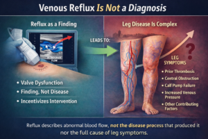
Varicose veins begin with valvular incompetence, which means the vein’s one-way valves no longer close properly. Instead of forming a tight seal, they leak—allowing reflux, the backward flow of blood that should only move upward toward the heart. This failure disrupts normal venous circulation and sets off a cascade of harmful hemodynamic changes.
When valves become incompetent:
Gravity pulls blood downward.
Because the valves cannot stop backward flow, blood slides down the vein under the constant force of gravity, especially when standing.Hydrostatic pressure in the vein rises.
Hydrostatic pressure refers to the pressure exerted by a column of fluid—in this case, blood—due to gravity. As more blood pools in the lower segments of the vein, the weight of this blood column increases the internal pressure.The vein wall dilates from increased pressure.
Vein walls are flexible, so rising hydrostatic pressure stretches and widens them. This dilation makes the vein more tortuous and visible at the surface.A dilated vein further impairs valve closure, worsening reflux.
Once the vein expands, the valve leaflets are pulled apart and can no longer meet in the middle. This makes reflux more severe and continuous, accelerating the progression of varicose veins.
This creates a self-perpetuating loop.
Key Hemodynamic Change:
Reflux + Venous hypertension → Vein wall dilation → More reflux
This cycle explains why varicose veins gradually enlarge and spread.
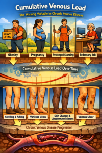
Venous Hypertension: The Engine Behind Symptoms
Chronic venous hypertension refers to abnormally high pressure inside the veins, and it is the main force behind most symptoms and complications of varicose vein disease. In hemodynamic terms, hypertension means that the venous system is carrying more pressure than it is designed to handle. This elevated pressure disrupts normal blood flow, causes fluid to leak into surrounding tissues, and forces the vein walls to stretch and weaken over time. As venous hypertension persists, it becomes the driving mechanism that leads to swelling, pain, inflammation, skin changes, and eventually venous ulcers.
Why does venous pressure rise?
Reflux increases the volume of blood pooling in the limb.
The calf muscle pump becomes inefficient due to overloaded veins.
Perforator incompetence allows deep venous pressure to transmit into the superficial system.
Consequences of high venous pressure
Heaviness and aching
Leg swelling
Inflammation in the vein wall
Skin discoloration (blood component leakage in the skin)
Dermatitis ( like eczema)
Venous ulceration in advanced disease ( sores on the leg)
Hemodynamically, the energy loss from turbulent, retrograde flow contributes to further venous failure.
The Role of Vein Wall Biology in Hemodynamics
Hemodynamics and vein biology interact intimately:
High pressure stretches the vein.
Stretch causes remodeling of collagen and elastin.
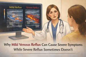
The vein becomes less elastic and more distensible.
Valve leaflets can no longer meet—worsening reflux.
This turns what begins as a mechanical problem into a structural one. Initially, the issue is purely mechanical: the valves fail to close properly, allowing blood to reflux. But over time, the abnormally high pressure and repetitive stretching caused by this dysfunctional flow begin to alter the structure of the vein itself. The vein wall remodels—its collagen and elastin fibers weaken, the vessel becomes more distensible, and the valve leaflets lose their ability to meet in the center. As a result, the vein no longer has the physical integrity to function normally, even if the mechanical forces were corrected. What started as a simple failure of valve movement evolves into a permanent change in the anatomy of the vein, reinforcing and worsening the cycle of reflux and dilation.
5. Hemodynamics of Perforator Veins: The Hidden Players
Perforator veins are small veins that connect the superficial veins near the skin to the deeper veins inside the leg. Their job is to move blood from the outer system into the deep system, where the leg muscles can pump it efficiently back to the heart. They contain one-way valves that make sure blood flows in the right direction. When perforator veins work properly, they help keep pressure low in the superficial veins and prevent swelling or vein enlargement. But if their valves fail, blood flows backward from the deep veins into the superficial ones, creating high pressure that can stretch veins, damage the skin, and contribute to varicose veins and leg ulcers.
Perforator veins normally direct blood from superficial → deep.
In varicose veins, incompetent perforators reverse this gradient:
Deep system pressure forces blood into superficial veins.
Localized high pressure damages surrounding tissues.
This is a key mechanism in venous stasis skin changes and ulcer formation.
When perforators fail, the entire hemodynamic load of the leg becomes unstable.
Why Varicose Veins Often Appear in Specific Locations
Venous junctions are key connection points where major superficial veins meet the deep venous system. The two most important are the saphenofemoral junction (SFJ) in the groin and the saphenopopliteal junction (SPJ) behind the knee. These junctions contain valves that normally prevent blood from flowing backward into the superficial veins. When reflux occurs at the SFJ, the great saphenous vein becomes overloaded, leading to varicose veins along the inner thigh. When reflux occurs at the SPJ, the small saphenous vein is affected, causing varicosities in the calf. Because these are high-flow areas, any valve failure rapidly increases pressure in the connected superficial segments, which are thin-walled and stretch easily. As a result, the superficial veins closest to the faulty junction dilate first and become visibly varicosities.
Hemodynamics explain typical patterns:
Saphenofemoral junction (SFJ) reflux → great saphenous varicosities along the inner thigh
Saphenopopliteal junction (SPJ) reflux → small saphenous varicosities in the calf
High-flow superficial segments experience the greatest pressure and dilate first.
Anatomical variations also affect flow distribution, making some people more prone to varicosities.
Standing vs Walking: How Hemodynamics Change Through the Day
Standing still
Venous pressure peaks (up to 80–100 mmHg in the ankle). For perspective, 80–100 mmHg of pressure in the ankle is similar to the pressure inside a car tire. It’s far higher than what veins are designed to handle, which is why standing still for long periods can cause discomfort, heaviness, and swelling in people with venous disease.
Reflux is greatest.
Symptoms worsen.
Walking
Calf pump reduces venous pressure significantly (to < 30 mmHg in healthy legs).
But in varicose veins, the pressure does not drop normally due to reflux → the hallmark of failed venous hemodynamics.
This explains why patients feel relief when walking but heaviness after standing.
Modern Treatments Target Hemodynamics—Not Just Appearance
Every effective treatment focuses on interrupting reflux and reducing venous hypertension, such as:
Endovenous laser or radiofrequency ablation: closes refluxing veins
Foam sclerotherapy: obliterates poorly functioning segments
Phlebectomy: removes bulging tributaries
Compression therapy: reduces local pressure and improves venous return
The goal is to restore normal flow patterns or eliminate the pathways that cause reflux.
Conclusion: Hemodynamics Are the Heart of Varicose Vein Disease
Varicose veins aren’t just enlarged veins—they are the result of a complex hemodynamic failure involving:
Valve incompetence
Reflux
Venous hypertension
Vein wall remodeling
Disturbed flow between superficial and deep systems
Understanding the hemodynamics gives clinicians a roadmap for effective diagnosis and treatment—and helps patients understand why the condition progresses.